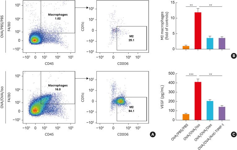
Fig. 7. The effects of anti-TIMP-1 antibody treatment on mouse macrophages. (A) The percentage of macrophage count (CD45+F4/80+ cells) and M2 macrophages (CD11c−CD206+ macrophages) measured by flow cytometry. (B) The degree of M2 macrophage count in lung tissue. (C) The levels of VEGF in the BALF (n = 5 for each group). Data are presented as mean ± standard deviation.
BALF, bronchoalveolar lavage fluid; Dex, dexamethasone; OVA, ovalbumin; TIMP-1, tissue inhibitor of metalloproteinase-1; VEGF, vascular endothelial growth factor; PBS, phosphate buffered saline.
**P < 0.01 and ***P < 0.001 by one-way analysis of variance with the Bonferroni post hoc test.

