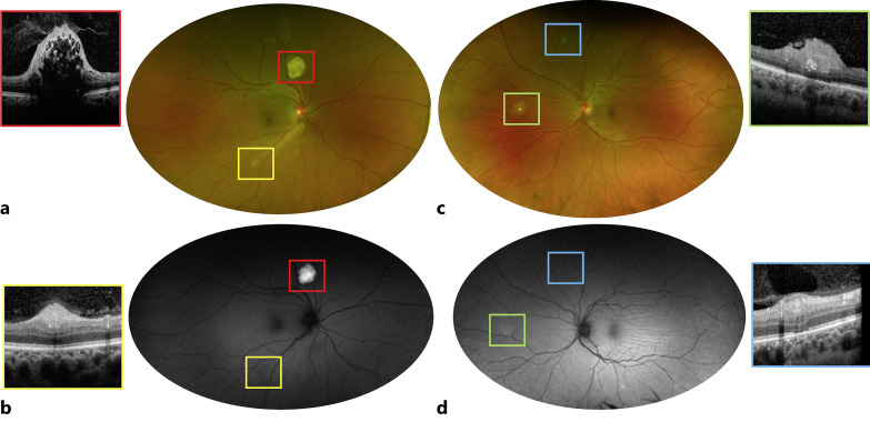
Fig. 1.
Bilateral retinal findings in a 43-year-old female patient with TSC. Fundus photo of the right eye (a) reveals a calcified, mulberry-shaped type 3 RAH superior to the nerve (red square) with hyper-autofluorescence (b) compatible with internal calcifications. Multiple, opaque, smaller lesions are visible along the inferior arcade (a), type 1 RAHs limited to the RNFL (yellow square) and iso-autofluorescent (b). c, d These solitary and flat type 1 RAHs are also present in the left eye (blue square); a nasal type 2 RAH is present in the left eye (c, green square) with elevation and some degree of retina traction (green square) and mild hyper-autofluorescence (d, green square).

