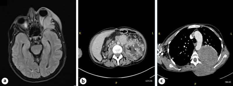
Fig. 2.
Systemic imaging studies of a 43-year-old female with TSC. MRI brain and orbit (a) shows left eye proptosis due to aggressive solid enhancing heterogenous infiltrative lesion arising from the lateral orbital wall with extensive bone destruction of left medial cranial fossa and extending into the orbital canal, displacing the left optic nerve medially. There is compressive mass effect on the left inferior rectus muscle as well as the left superior rectus muscle. Her CT abdomen (b) shows left renal enlargement, angiomyolipoma, and polycystic kidney disease. Her chest CT (c) shows a large left lung mass that on incisional biopsy was positive for a morphological diagnosis of a PEComa.

