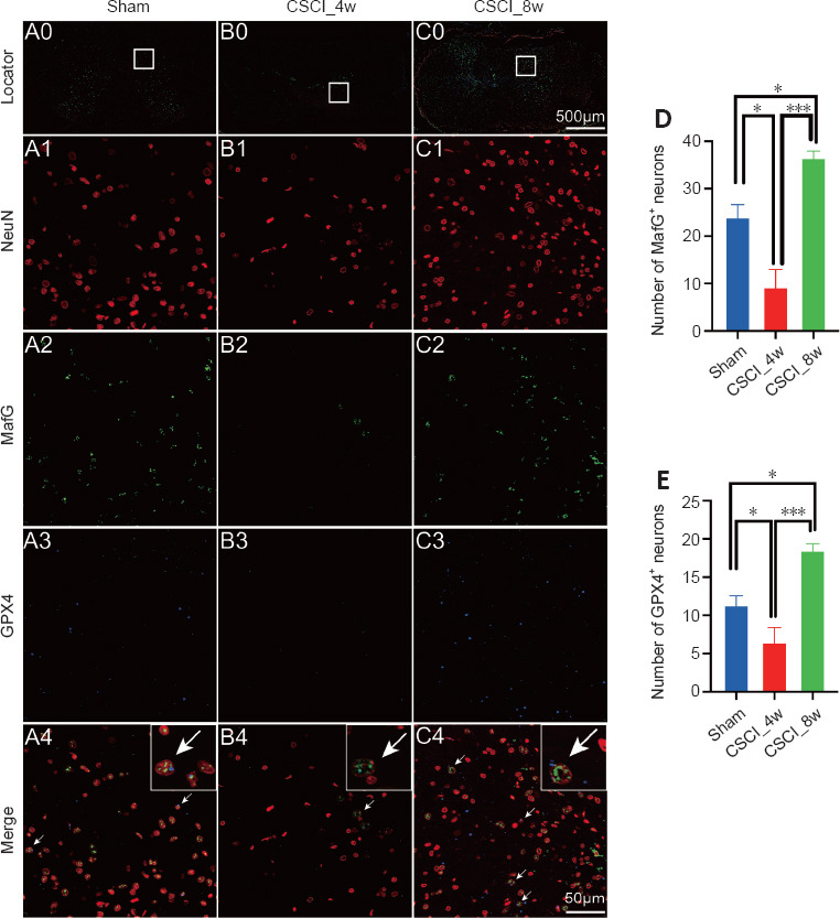
Figure 6.

IF of MafG and GPX4 in neurons after chronic CSCI.
(A0–C0) Overview images indicating the location of the magnified IF images. (A1–3 to C1–3) Magnified IF images stained with NeuN (A1–C1, red), MafG (A2–C2, green), GPX4 (A3–C3, blue) and merged channels (A4–C4). The white arrows indicate NeuN+/MafG+/GPX4+ triple-labeled cells. NeuN and GPX4 are located in the cytoplasm of neuronal cells, whereas MafG primarily resides in the nuclei. (D, E) Numbers of MafG+ and GPX4+ neurons in each group. *P < 0.05, ***P < 0.001 (Student’s t-test). Sample sizes: sham, n = 4; CSCI_4w, n = 4; and CSCI_8w, n = 4. Data are presented as the mean ± standard deviation. CSCI: Compressive spinal cord injury; IF: immunofluorescence; MafG: MAF BZIP transcription factor G; GPX4: glutathione peroxidase 4.
