
Figure 3.
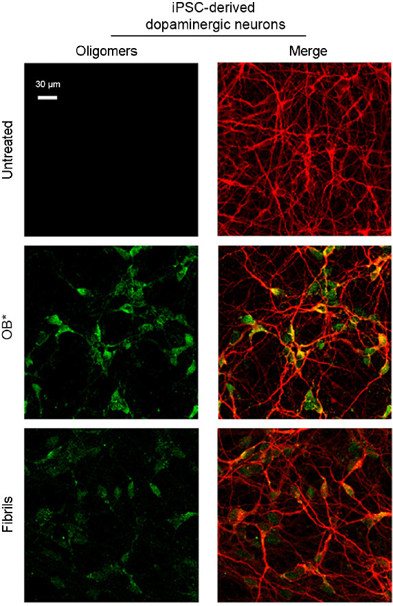
Representative confocal scanning microscope images showing human induced pluripotent stem cell (iPSC)-derived dopaminergic neurons treated for 24 hours with type B* oligomers (OB*) and fibrils of α-synuclein at 0.3 μM.
Red and green fluorescence indicates mouse anti-MAP-2 antibodies and the A11-positive prefibrillar oligomers, respectively. Images adapted and reprinted from Cascella et al. (2021), licensed under Creative Commons Attribution 4.0 International Public License (CC BY 4.0, https://creativecommons.org/licenses/ by/4.0/).