
Figure 6.
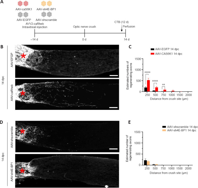
S6K1 activation, but not the knockdown of 4E-BP1, is sufficient to promote axon regeneration.
(A) AAV-caS6K1 and AAV-sh4E-BP1 were administrated intravitreally to mice 2 weeks before ONC. CTB was intravitreally injected 12 days after ONC and optic nerves were harvested on day 14 after ONC. (B) Expression of caS6K1 induced axon regeneration. Images of optic nerve sections show regenerating axons labeled by anterograde transport of CTB on day 14 after optic nerve crush from AAV-EGFP- and AAV-caS6K1-injected eyes. (C) Quantification of regenerating axons 14 days post-crush from AAV-EGFP- and AAV-caS6K1-injected eyes at different distances distal from the crushing site (mean ± SD, n = 6–9). (D) Knockdown of 4E-BP1 did not induce axon regeneration. Images of optic nerve sections show regenerating axons labeled by anterograde transport of CTB on day 14 after ONC from AAV-shscramble and AAV-sh4E-BP1-injected eyes. Red asterisk indicates crush site. Scale bars: 100 µm (B, D). (E) Quantification of regenerating axons 14 days post-crush from AAV-shscramble- and AAV-sh4E-BP1-injected eyes at different distances from the crushing site (means ± SD, n = 6). **P < 0.01, ****P < 0.0001 (two-way analysis of variance with Bonferroni’s post hoc test). 4E-BP1: Mammalian target of rapamycin complex 1 downstream effector; AAV: adeno-associated virus; caS6K1: constitutively active S6K1; CTB: cholera toxin B subunit; dpc: days post crush; EGFP: enhanced green fluorescent protein; ONC: optic nerve crush; RGC: retinal ganglion cell; S6K1: mammalian target of rapamycin complex 1 downstream effector.