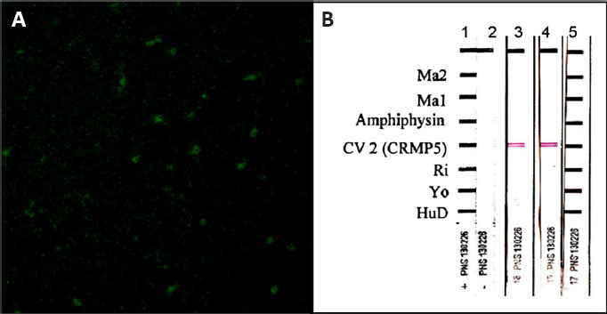
Figure 2.

Patient’s serum antibody reactivity.
(A) A pattern suggesting anti-CV2 positivity (cytoplasmic staining with nuclear sparing) obtained by indirect immunofluorescence performed with our patient’s serum on primate cerebellum (Inova Diagnostics, San Diego); the estimated titer was 1:500. (B) Subsequent immunoblot verification (Ravo Diagnostika) of anti-CV2 isolated presence in serum obtained from the proband in two separate occasions (lanes 3 and 4); serum dilution 1:2000. Reproduced with permission from Aliprandi et al. (2015).
