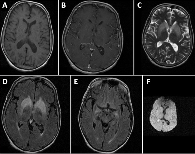
Figure 3.

MRI of a patient with CV2/CRMP5 antibody-associated paraneoplastic neurological syndrome.
(A, B) T1-weighted plain and contrast MRI results are normal. (C, D) T2-weighted and FLAIR images show bilateral symmetrical basal ganglia hyperintensities. (E) FLAIR image shows bilateral mesial temporal hyperintensities. (F) Diffusion weighted image is normal. Reproduced with permission from Maramattom (2013). CRPM5: Collapsin response mediator protein 5; MRI: magnetic resonance imaging.
