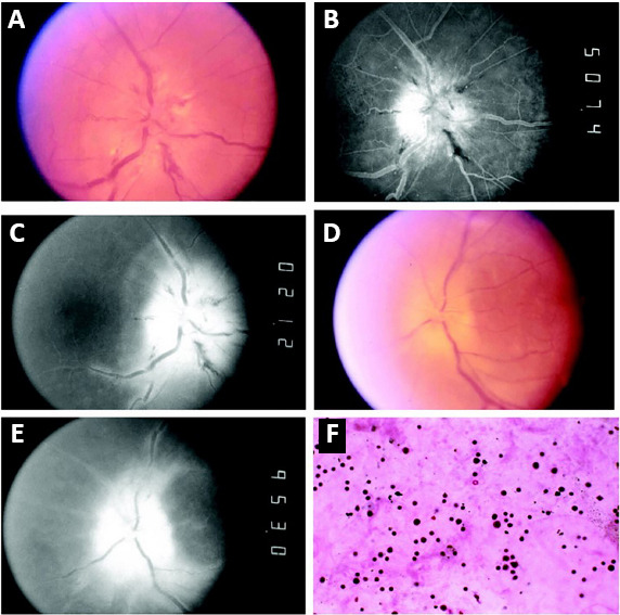
Figure 5.

Optic disc photographs, fluorescein images, and vitreous biopsy.
(A) Right eye shows severe acute optic disc swelling with tortuous vessels, cotton wool spots, and nerve fiber layer hemorrhages. (B) Late arterial fluorescein image shows dye leakage on disc surface, and (C) late phase shows dye persistence at the disc and perivenous fluorescence indicates vascular incompetence. (D, E) Left eye shows chronic disc swelling, (D) optic nerve atrophy with dye leakage and (E) vascular incompetence remote from the disc. (F) Pleomorphic cellularity consistent with reactive lymphocytosis in the vitreous humor of the right eye (hematoxylin and eosin, original magnification 400×). Reproduced with permission from Cross et al. (2003).
