
Figure 5.
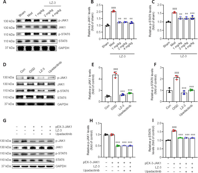
LZ-3 inhibits ischemia- or hypoxia-induced JAK1-STAT6 signaling.
(A) Representative western blot showing p-JAK1 and p-STAT6 expression in the cortex on day 3 after reperfusion. (B, C) Quantification of p-JAK1 and p-STAT6 expression based on western blotting (n = 3/group). ###P < 0.001, vs. sham group; **P < 0.01, ***P < 0.001, vs. model group. (D) Representative western blot showing p-JAK1 and p-STAT6 expression in BV2 cells treated with LZ-3 or upadacitinib after 24 hours of re-oxygenation after OGD. (E, F) Quantification of p-JAK1 and p-STAT6 expression based on western blotting (n = 3). (G) Representative western blot showing p-JAK1 and p-STAT6 expression in BV2 cells overexpressing JAK1 and treated with LZ-3 or upadacitinib after 24 hours of re-oxygenation after OGD. (H, I) Quantification of p-JAK1 and p-STAT6 expression based on western blotting (n = 3). ###P < 0.001, vs. control group; ***P < 0.001, vs. OGD group. Data are expressed as mean ± SD and were analyzed by one-way analysis of variance followed by Tukey’s post hoc test. Con: Control group; GAPDH: glyceraldehyde-3-phosphate dehydrogenase; JAK1: Janus kinase 1; Mod: model group; OGD: oxygen-glucose deprivation; pEX-3-JAK1: peroxisomal biogenesis factor 3-Janus kinase 1; p-JAK1: phospho-Janus kinase 1; p-STAT6: phospho-signal transducer and activator of transcription; STAT6: signal transducer and activator of transcription.