
Figure 4.
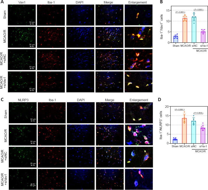
Vav1 knockdown represses the activation of microglia and NLRP3 inflammasome in the ischemic penumbra of MCAO/R rats.
(A) Double immunofluorescence staining for Vav1 (FITC, green) and Iba-1 (Cy3, red) in the ischemic penumbra area in MCAO/R rats. MCAO/R rats showed enhanced expression of Vav1 and Iba-1 compared with the sham group, but inhibition of Vav1 expression decreased the levels of both proteins. (B) Quantification of Vav1/Iba-1 double positive cells. (C) Double immunofluorescence staining for NLRP3 (FITC, green) and Iba-1 (Cy3, red) in the ischemic penumbra area in MCAO/R rats. MCAO/R rats showed enhanced expression of Iba-1 and NLRP3 compared with the sham group, but inhibition of Vav1 expression decreased the levels of both proteins. Nuclei were stained with DAPI (blue). Scale bars: 50 μm. (D) Quantification of NLRP3 and Iba-1 double positive cells. Data are expressed as the mean ± SD (n = 6), and were analyzed by one-way analysis of variance followed by Tukey’s multiple comparisons test. Cy3: Cyanine 3; DAPI: 4′,6-diamidino-2-phenylindole; FITC: fluorescein isothiocyanate; MCAO/R: middle cerebral artery occlusion/reperfusion; NLRP3: NOD-like receptor pyrin 3; Vav1: Vav guanine nucleotide exchange factor 1.