
Figure 4: Dissection of the sinoatrial (SAN) node.
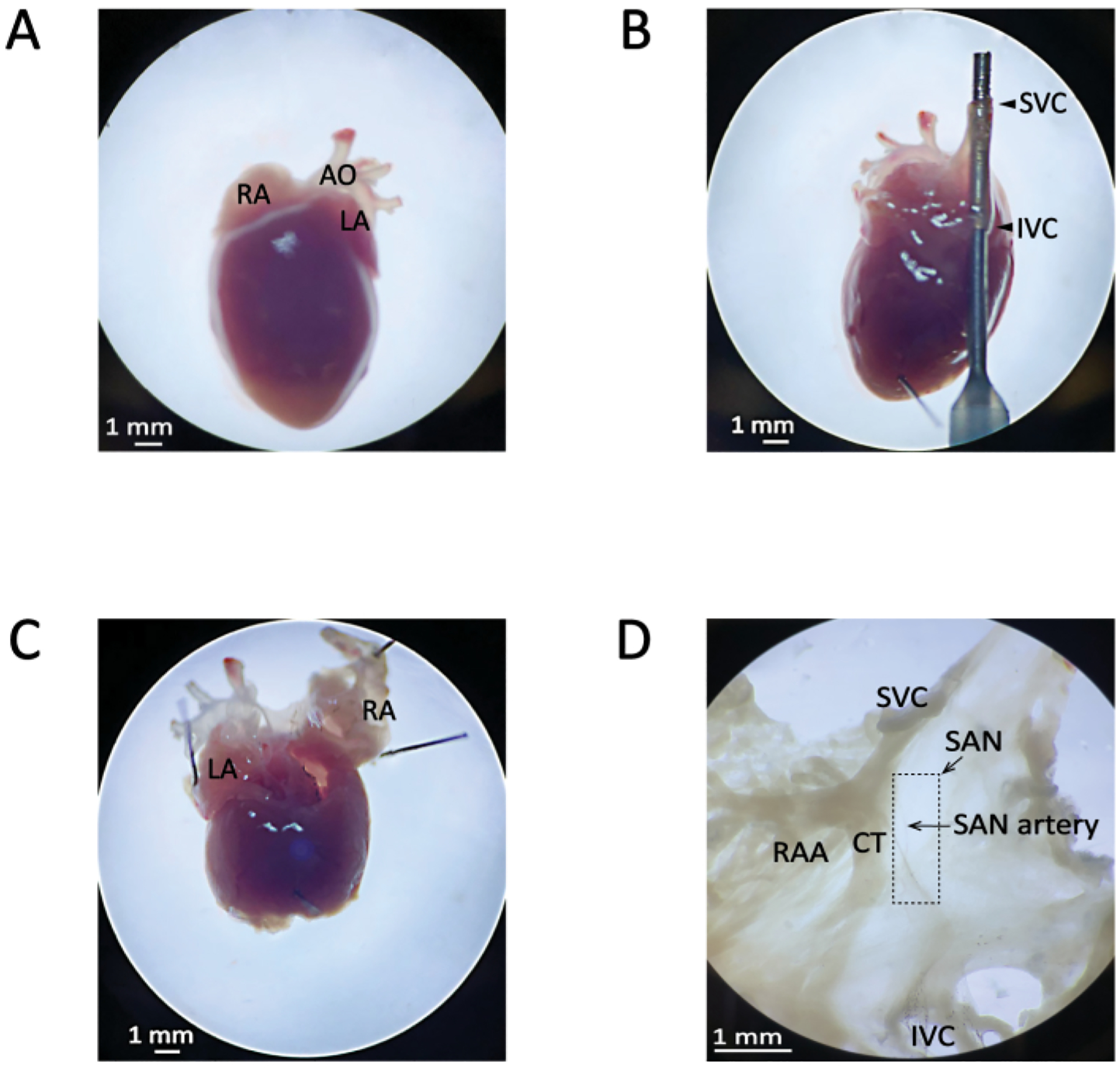
(A) The appearance of the heart in the Petri dish following removal from the body. (B) Insertion of the syringe needle through the inferior vena cava (IVC) and superior vena cava (SVC) of the right atrium. The pin in the apex of the heart is also shown. (C) Excision of the apex (i.e., the bottom half) of the heart to release the blood. The pins in the atrial appendages are also shown. (D) The final appearance of the SAN region of the right atrium at the end of dissection. The boxed region corresponds to the approximate location of the SAN. The SAN artery can also be faintly seen coursing through the SAN in a vertical orientation. The Abbreviations: AO, aorta; CT, crista terminalis; IVC, inferior vena cava; LA, left atrium; RA, right atrium; RAA, right atrial appendage; SAN, sinoatrial node; SVC, superior vena cava.