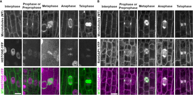
Figure 2.
Chromosome marker HISTONE1.1-YFP and nuclear envelope marker RANGAP1-YFP with CFP-TUBULIN during the cell cycle. Microtubules from the abaxial side of maize leaves in regions with symmetrically dividing cells were imaged with CFP-TUBULIN (top row). In the merged images, microtubules are colored green, while HISTONE1.1-YFP or RANGAP1-YFP are colored magenta. (A) Chromosomes are labeled with HISTONE1.1-YFP (middle row). (B) RANGAP1-YFP labels the cell periphery, the nuclear envelope during interphase and preprophase/prophase, and localizes close to the PPB during prophase/preprophase. During mitosis, RANGAP1-YFP evenly labels all microtubule structures but is not at the division site. Preprophase/prophase RANGAP1-YFP and CFP-TUBULIN images are maximum-projections of Z stacks covering 7.5 microns to more clearly illustrate RANGAP1-YFP localization near the PPB. Scale bar is 10µm, all images are the same size.

