Figure 1.
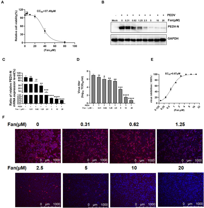
The cellular toxicity and anti-PEDV activity of Fan in IPEC-J2 cell cultures. (A) IPEC-J2 cells were treated with various concentrations of Fan at 37°C for 48 h. Cell viability was evaluated using CCK-8 assays. (B–D) Fan at various concentrations was used to treat cells for 1 h before PEDV GD/HZ/2016 infection (0.1 MOI), and the cells were then treated with various Fan concentrations for 24 h. At 24 hpi, supernatants and intact cells were collected. (B) Western blotting assessment of PEDV N protein levels. (C) QRT-PCR quantification of PEDV N mRNA levels. (D) TCID50 assay to determine the viral titers. (E) Assessment of the EC50 of Fan toward PEDV infection. (F) Effect of Fan on the inhibition of PEDV analyzed using IFA. *P < 0.05; **P < 0.01; ***P < 0.001; and ****P < 0.0001 indicate significant differences vs. the control group.

