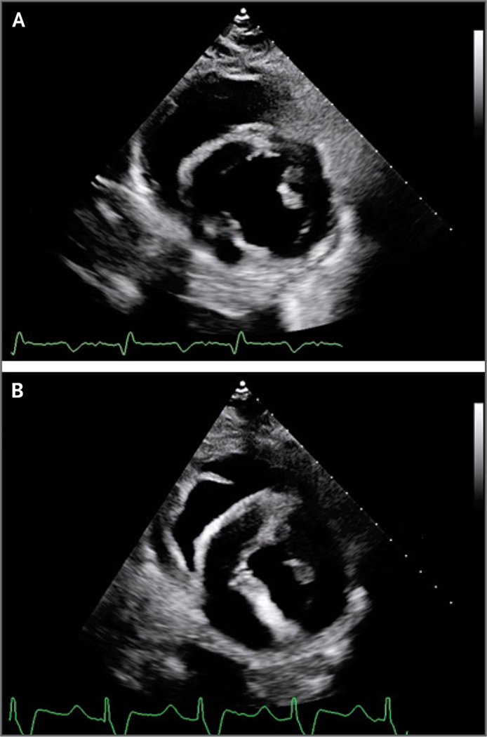
Figure 2. Transthoracic Echocardiography.
Transthoracic echocardiography of the transplanted xenograft obtained on day 19 after transplantation (Panel A, parasternal short axis) showed normal xenograft function, normal global longitudinal strain, and normal left ventricular posterior wall thickness in end diastole. Transthoracic echocardiography performed on day 49 (Panel B), before cannulation for venoarterial extracorporeal membrane oxygenation (ECMO), showed preserved systolic function, less negative (i.e., more abnormal) global longitudinal strain, and increased thickness of the left ventricular posterior wall in end diastole.

