
Figure 4. Histologic Assessments.
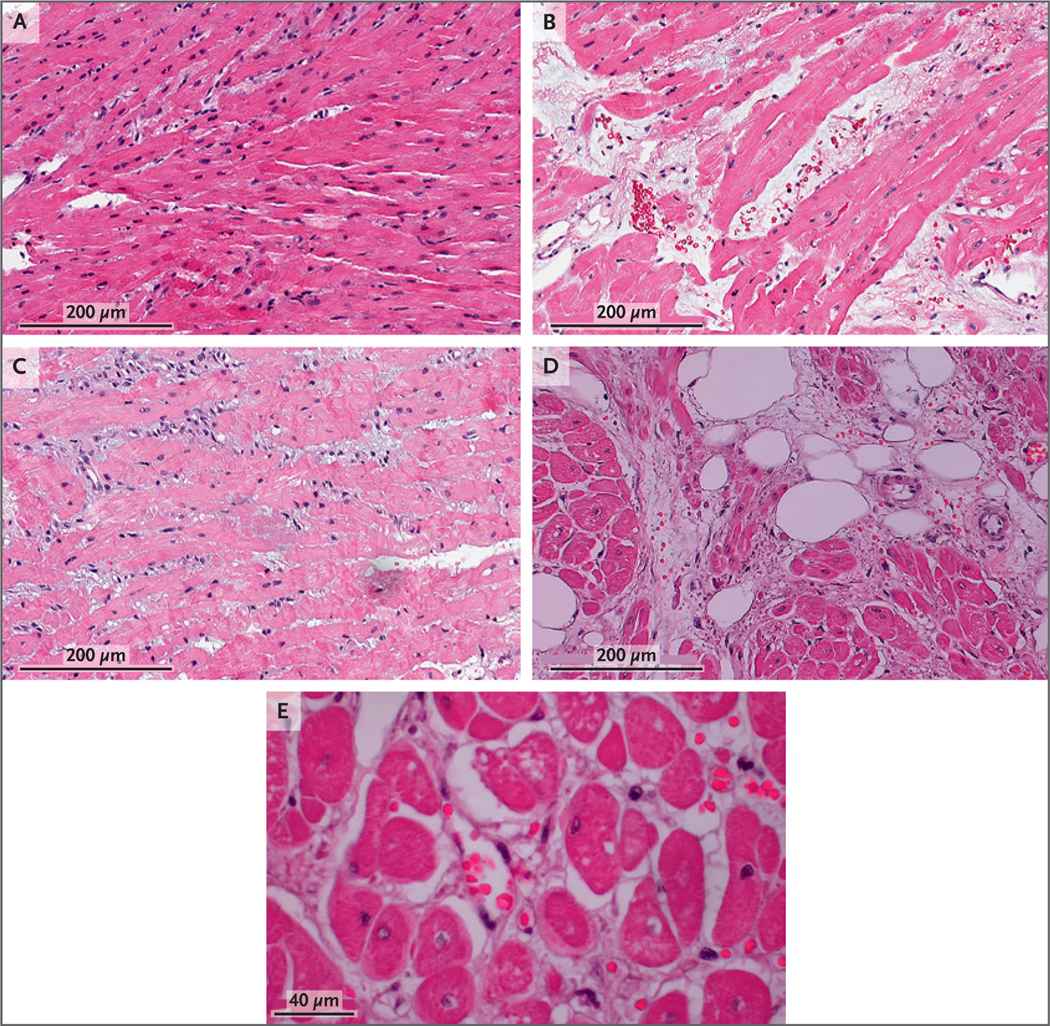
The first endomyocardial biopsy (Panel A, day 34 after transplantation) showed normal histologic characteristics. The second endomyocardial biopsy (Panel B, day 50) showed interstitial red-cell extravasation, fragmentation, and edema without cellular infiltrate or intravascular thrombosis. The third endomyocardial biopsy (Panel C, day 56) showed some resolution of interstitial edema and red-cell extravasation but also showed evidence of necrotic myocytes. The septum at autopsy (Panels D and E, day 60) showed interstitial edema and red-cell extravasation, myocardial necrosis, scant fibrosis, and central nuclei.