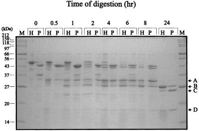
FIG. 2.
Tryptic digestion of ParB and His-ParB. ParB (lanes P) and His-ParB (lanes H) were treated with trypsin at a protein/protease ratio of 1,000:1 (wt/wt) at 20°C for the indicated times. Digestion was stopped with 1% acetic acid. Proteolytic fragments were separated by electrophoresis in SDS–15% polyacrylamide gels and were visualized with Coomassie blue. Undigested ParB and His-ParB migrate with 44- and 50-kDa proteins, respectively. The arrows at the right indicate the major tryptic fragments identified in Table 3. Lane M, size markers.

