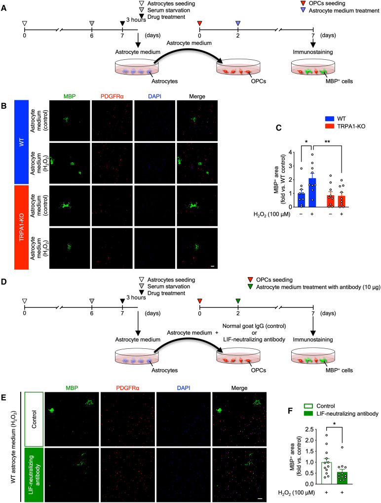
Fig. 9. H2O2-elicited TRPA1-mediated astrocytic LIF promotes myelination of cultured OPCs.
(A) Experimental time course for in vitro medium transfer experiments from astrocytes to OPCs. (B and C) Representative images of immunostaining with anti-MBP antibody (green), anti-PDGFRα antibody (red), and 4′,6-diamidino-2-phenylindole (DAPI) (blue) in primary OPC cultures treated with astrocyte medium (B) and summarized data for the percentage of MBP-positive surface areas (C). (D) Experimental time course for in vitro medium transfer experiments with a LIF-neutralizing antibody. (E and F) Representative images of immunostaining with anti-MBP antibody (green), anti-PDGFRα antibody (red), and DAPI (blue) in primary OPC cultures treated with H2O2-treated WT astrocyte medium with LIF-neutralizing antibody or control antibody (E) and summarized data for the percentage of MBP-positive surface areas (F). Values are means ± SEM. Scale bars, 100 μm. (C) n = 9; (F) n = 12. *P < 0.05 and **P < 0.01 for two-way ANOVA with Bonferroni’s post hoc test (C). *P < 0.05 for two-tailed unpaired Student’s t test (F).

