
Fig. 1 |. Atherosclerotic plaque formation and vascular inflammation in mice lacking platelet sGC.
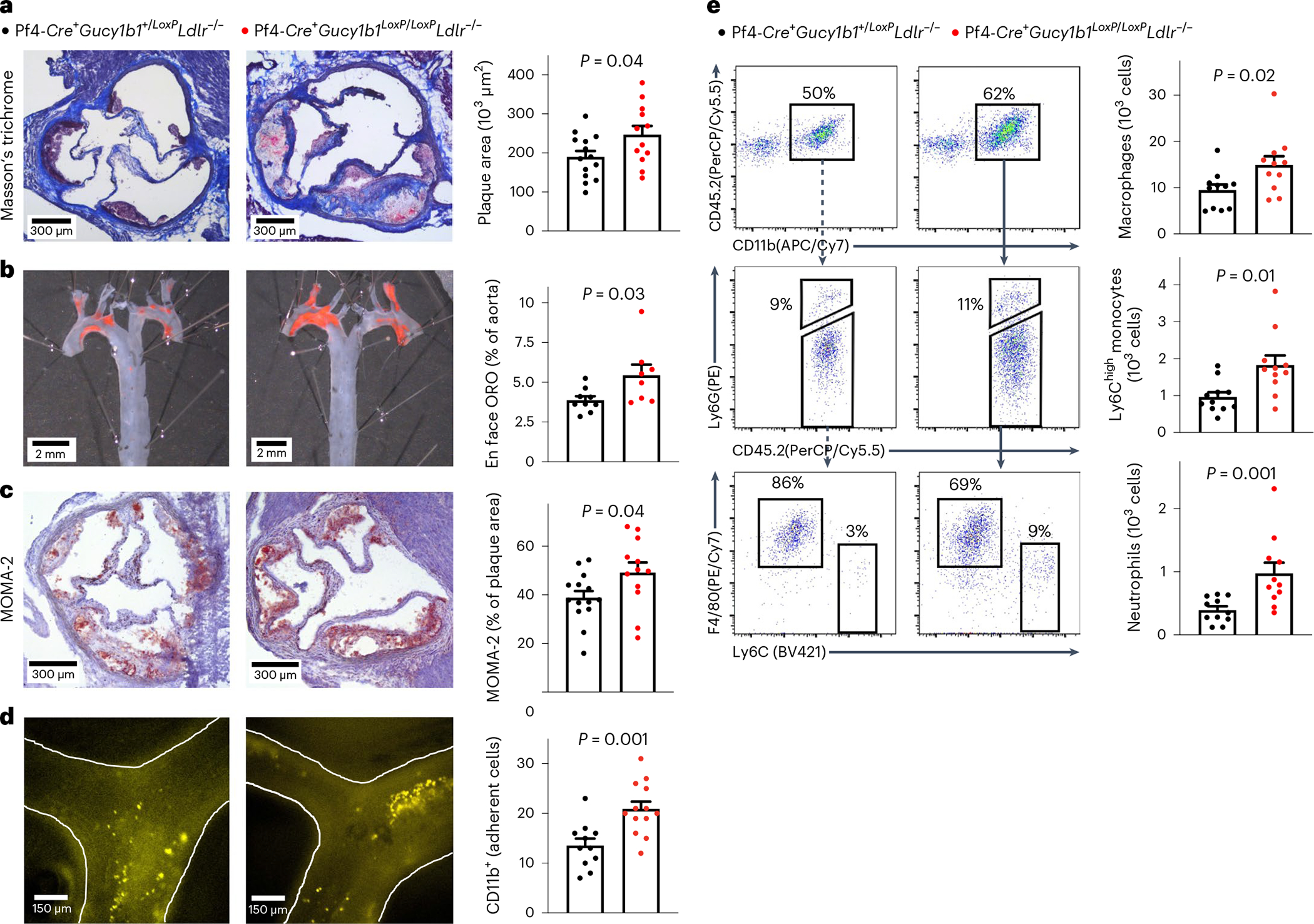
a–c, Atherosclerotic plaque formation as assessed by aortic root histology (a), aortic en face ORO staining (b), and monocyte and macrophage content (c) in 12 (b: 8) Pf4-Cre+Gucy1b1LoxP/LoxPLdlr−/− mice compared to 14 (b: 9) Pf4-Cre+Gucy1b1+/LoxPLdlr−/− mice that were fed a Western diet for 10 weeks. Two-sided unpaired t-test. d, Leukocyte adhesion to atherosclerotic plaques in n = 13 Pf4-Cre+Gucy1b1LoxP/LoxPLdlr−/− mice compared to n = 11 Pf4-Cr e+Gucy1b1+/LoxPLdlr−/− mice that were fed a Western diet for 6 weeks to induce atherosclerotic plaque formation. Two-sided unpaired t-test. e, Quantification of vascular inflammation by flow cytometry analysis of aortic cell suspensions of Pf4-Cre+Gucy1b1LoxP/LoxPLdlr−/− mice compared to Pf4-Cre+Gucy1b1+/LoxPLdlr−/− mice (n = 11 per group). Two-sided Mann–Whitney U-test. Each symbol represents one independent animal. Data are the mean ± s.e.m.