
Figure 4:
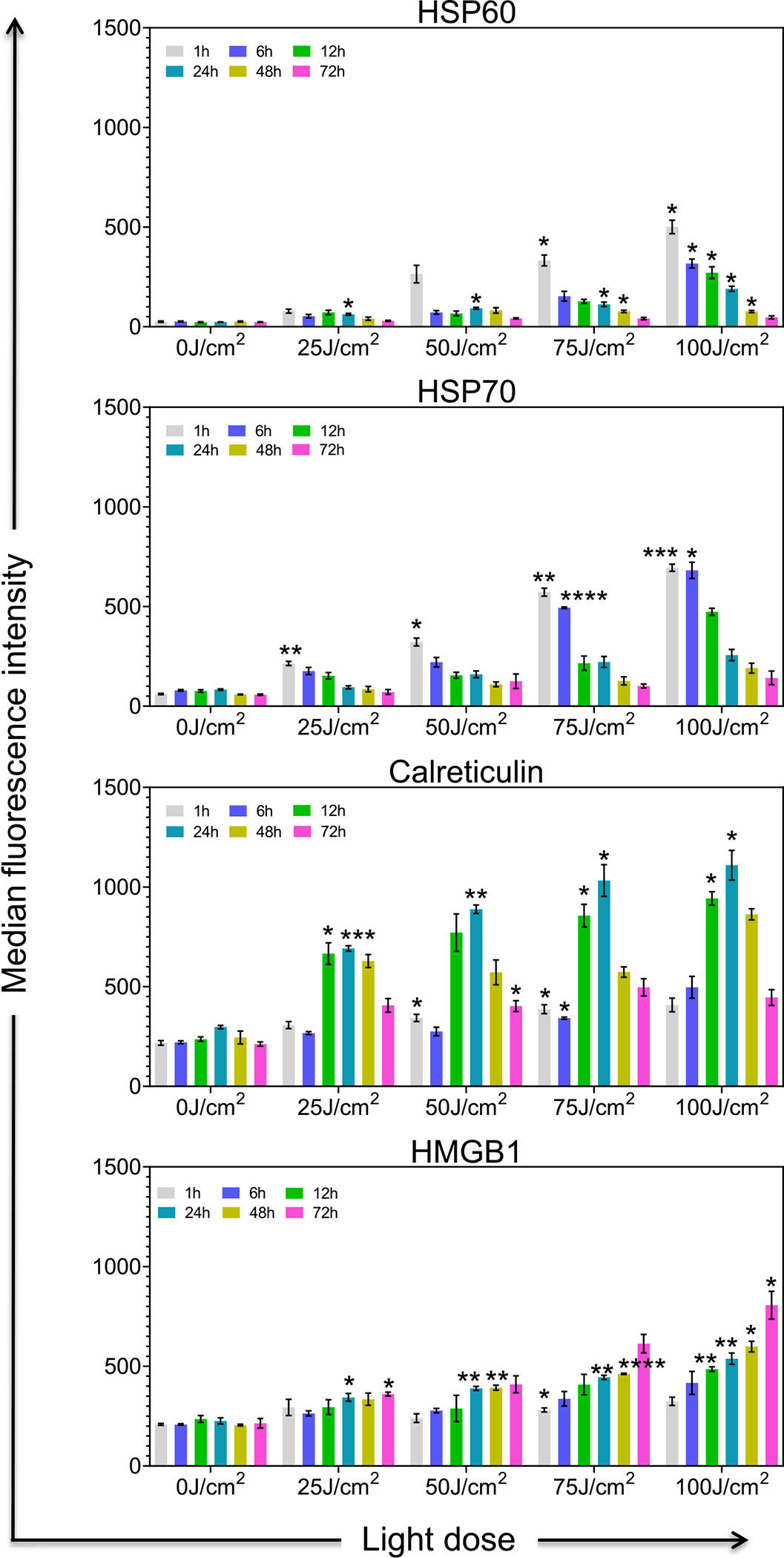
Expression of TR-PINs induced biological markers of immunogenic cell death in Panc spheroid cultures. NIR activation of TR-PINs induces cell surface exposure of Hsp60, Hsp70, Calreticulin, and the intracellular expression of HMGB1 in Panc (MIA PaCa-2 and PCAF) spheroid cultures in a light dose and time-dependent manner. Data are representative of three independent experiments done in duplicates. Expression levels of Hsp60, Hsp70, Calreticulin, and HMGB1 were determined by flow cytometry calculated as the median fluorescence intensity (MFI) after subtraction of the isotype controls MFI at 1, 6, 12, 24, 48, and 72 h after NIR activation of TR-PINs. Graphs with error bars indicate mean ± SEM from three independent experiments. Statistical significance was determined by a one-way ANOVA and Tukey’s posthoc test. Asterisks denote statistical significance (*P < 0.05, **P < 0.005, ***P < 0.0005, ****P < 0.00005). The NIR photodynamic activation regimen used was 690 nm light irradiation with 25 or 50 or 75 or 100 J/cm2 at 150 mW/cm2. Two hundred and fifty Newton-meters of TR-PINs (BPD-PC equivalent) were used.