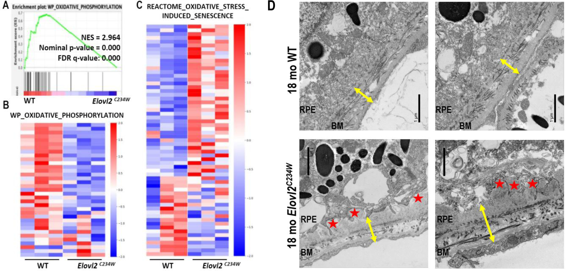
Figure 5. Metabolic and structural changes in ELOVL2 deficient animals.

Gene set enrichment analysis (GSEA) was performed on RNA-Seq data from the retina of Elovl2C234W and WT mice. A. “Oxidative Phosphorylation” pathway is significantly downregulated, as shown on the enrichment plot. B. Gene sets “Oxidative Phosphorylation” and C. “Oxidative Stress Induced Senescence’’ with genes ranked by WT/Elovl2C234W in mean log2Fold Change of expression level. Color bars indicate row Z-score. In the analysis, we used 4-month-old mice (N=4 for each genotype) and Molecular Signatures Database (MSigDB). D. EM analysis of Bruch’s Membrane (BM). Comparison of 18-month-old WT and Elovl2C234W RPE/BM surfaces show thickening and disorganization of BM (yellow arrows), including accumulation of basal lamina deposits (BLamD - red stars) and distorted RPE infoldings
