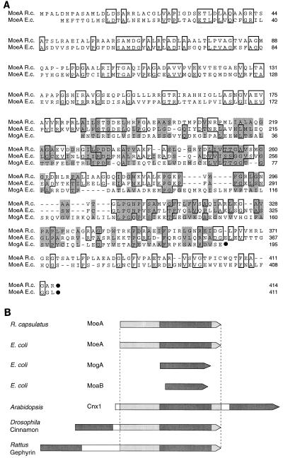
FIG. 2.
Alignment of the deduced amino acid sequence of R. capsulatus MoeA with E. coli MoeA and MogA. (A) Amino acid sequences were aligned for maximum matching by use of the CLUSTAL W program (44). The C-terminal ends of polypeptides are marked by black dots. Identical amino acids are boxed, and in addition, the similarity of amino acids between E. coli MogA, R. capsulatus MoeA, and E. coli MoeA is emphasized by shading. (B) Schematic overview of different bacterial and eukaryotic proteins showing similarities to E. coli MoeA and MogA. The E. coli MogA protein and MogA-like domains found in other proteins are indicated by dark gray shading. Protein domains showing similarities to E. coli MoeA are indicated by light gray shading.

