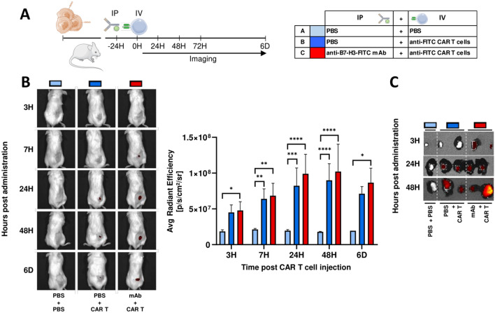
Fig. 5.
CAR T migration in vivo to 143B tumors. A Schematic representation of experimental design. PBS (blue) or 100 μg of anti-B7-H3-FITC mAb (red) is administrated by intraperitoneal injection (IP) (-24H) to 143B tumor-bearing NSG mice. Next day (0H), PBS (light blue = A) or 1 × 10⁶ DiR-labeled CAR T cells (dark blue = B, dark red = C) are intravenously injected (IV). B DiR-labeled CAR T cells homing is determinate by IVIS imaging system at indicated time points. Representative images are shown. Average radiant efficiency on tumor region is analyzed, and the mean ± SD of at least three mice is represented. *, P < 0.05; **, P < 0.01; ***, P < 0.001; ****, P < 0.0001 by two-way ANOVA with Tukey post hoc test. C Ex vivo imaging by IVIS of DiR-labeled CAR T cells on tumors collected at different time points

