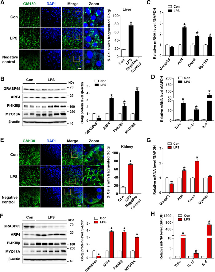
Fig. 2. LPS-induced endotoxemia promotes Golgi stress and inflammatory response in acute liver and kidney injury.
A Immunofluorescence staining of GM130 (green), a marker of Golgi morphology, in the liver tissues was assessed by confocal microscopy and quantification of cells with fragmented Golgi from each group is shown (n = 3). Scale bar, 20 µm. B Golgi functional and structural proteins (GRASP65, ARF4, P14KIIIβ, and MYO18A) were examined using western blot analysis in liver tissues and relative expression was shown over β-actin as loading control (n = 3–5). C Relative mRNA levels of Grasp65, Arf4, Creb3, and Myo18a were determined by real-time PCR analysis in liver tissues, and relative mRNA expression was normalized to that of GAPDH (n = 3–5). D Relative mRNA levels of pro-inflammatory cytokines (Tnfα, IL-1β, and IL-6) were determined by real-time PCR analysis in liver tissues and relative mRNA expression was normalized to that of GAPDH (n = 4). E–H As described above, the experiments in (A), (B), (C), (D) were similarly performed in kidney tissues. The data are presented as mean ± SEM. Two-tailed Student’s t-test was used, *p < 0.05 versus Control.

