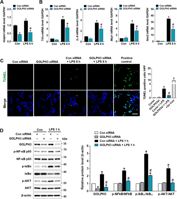
Fig. 5. Downregulation of GOLPH3 reduces LPS-induced inflammatory response, apoptosis, and AKT/NF-κB signaling activation in hepatocytes.
FL83B cells were transfected with Con siRNA or GOLPH3 siRNA for 24 h, and treated with LPS for 8 h. A, B After transfection, relative mRNA levels of Golph3 and pro-inflammatory mediators (Tnfα, IL-6, Mcp1, and Nos2) were examined using real-time PCR analysis. Relative mRNA expression was normalized to that of GAPDH (n = 3). C Representative images of TUNEL staining and the quantification of TUNEL-positive cells/HPF using Image J software (NIH) are shown. Recombinant DNase I (5 U/ml) treated cells were used as experimental positive control (n = 3). Scale bar, 50 µm. D After transfection, the cells were treated with LPS for 1 h, and the protein expression levels of GOLPH3, p-NF-κB p65, p-IκBα, p-AKT, and β-actin (as a loading control) were analyzed by western blot analysis (n = 3). The data are presented as mean ± SEM. One-way ANOVA, followed by Bonferroni’s multiple comparisons was used, *p < 0.05 versus Con siRNA alone, and #p < 0.05 vs LPS with Con siRNA.

