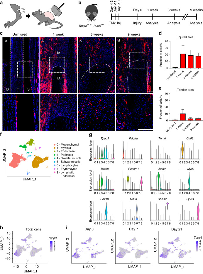
Fig. 1.
Tppp3+ tendon sheath progenitors expand at the heterotopic ossification (HO) induction site after Achilles tendon injury. a Schematic representation of HO induction, including complete Achilles tenotomy (left) and dorsal burn (right). b Tppp3ECE/+;R26RtdT animals were administered tamoxifen (TMx) for three continuous days, followed by a 10-day washout period before HO induction. Reporter activity was examined at 1, 3 and 9 weeks after injury. c tdT+ (Tppp3) cells (red) in the defect area were visualized using sagittal sections of the distal tenotomy site after injury. tdT+ cells were present outside of the tendon (epitenon) in the uninjured condition, and the cells expanded into the defect area of the tendon after injury. The dashed white line indicates the margins of the Achilles tendon, and the dashed white box in the upper panels is magnified in the lower panels (scale bars: 200 µm). D: deep; T: tendon; S: superficial, IA: injured area, TA: tendon area. d Fraction of tdT+ cells in the injured area. e Fraction of tdT+ cells in the residual tendon area (under dashed white line). f UMAP visualization of cell clusters from the HO induction site at 0, 7, and 21 d post-injury. g Violin plots showing the expression of markers for cell type identification. h UMAP of Tppp3 expression across all clusters (all time points together). i UMAP showing the expression of Tppp3 in cell clusters across time points. n = 3 animals per timepoint for histology; for scRNA-seq, n = 4 animals per timepoint; 3 678, 13 358 and 5 366 cells at timepoints 0, 7 and 21 d, respectively

