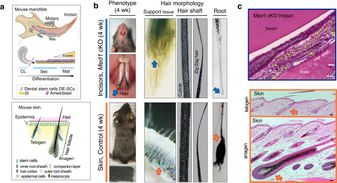
Fig. 1. Loss of Med1 in dental stem cells causes ectopic hair growth on incisors.
a Top, diagram representing normal dental epithelial differentiation and enamel formation in mouse mandible. CL cervical loop, Sec secretory stage, Mat maturation stage, DE-SCs dental epithelial stem cells, SI stratum intermedium. Bottom, hair regeneration by hair follicles under hair cycling regulation of resting telogen (left) and growing anagen (right) in skin. b Hair growth on Med1 cKO incisors at 4 weeks of age (top) is compared to normal hair in skin (bottom). Arrows show the root of hairs. c Top, HE sections of tissues emphasizing the atypical cell cluster (yellow triangles) surrounding hair shafts (yellow arrow) found between dentin and bone in Med1 cKO incisor (3 month). Bottom, HE sections for hair follicles at anagen (bottom) and telogen (top) phases supporting hair growth and regeneration in the skin (orange arrows). Bars = 50 μm. For b, c, representative images are shown. The diagram depicting the mouse mandible (brown colored) was created with BioRender.com, as were the ones shown in Figs. 2a, 3a, c, and 4a and Supplementary Figs. 1a and 4a.

