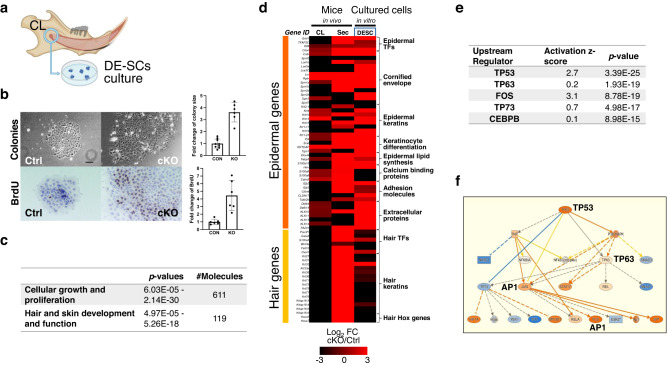
Fig. 4. Med1 deficiency directs DE-SCs towards epidermal fate in vitro.
a Generation of DE-SC culture from CL tissues. b Left, representative bright field images of DE-SC colonies and BrdU staining in Ctrl and Med1 cKO. Bar = 10 mm. Right, quantification of colony size and BrdU positive cells/colony are shown as fold changes (cKO/Ctrl) with standard deviations (error bars); in both comparisons (n = 6–9, t-test p < 0.01). c Biological processes associated with loss of Med1 in DE-SCs as identified by Ingenuity Pathway Analysis (IPA) on microarray gene expression data. d Heatmap showing differential gene expressions of epidermal and hair-related genes in Med1 cKO vs Ctrl incisor tissues (CL and Sec in vivo) and cultured Med1 cKO vs Ctrl DE-SCs (third lane); n = 3 for all samples and average values for each comparison are shown. e Upstream regulators responsible for biological process induced by loss of Med1 in DE-SCs as identified by IPA on microarray data as used in c. f Mechanistic network representation for TP53/63 pathways in induced in Med1 lacking DE-SC culture regulating epidermal fate driving AP-1 factors; upregulated genes are in orange and direct relationships are shown by solid lines. Reproducibility was confirmed by two independent cultures, and representative data are shown.

