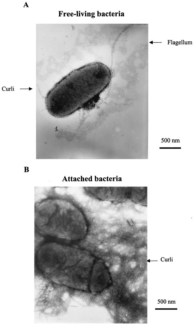
FIG. 2.
Electron micrographs of negatively stained bacteria of planktonic (A) or biofilm (B) cell suspensions of the motile PHL628 strain. The cells were grown in M63-mannitol medium, and biofilms were formed on plastic strips. The micrographs, corresponding to observations made after 24 h of culture, are representative of several microscopic observations performed at between 8 and 48 h of biofilm development.

