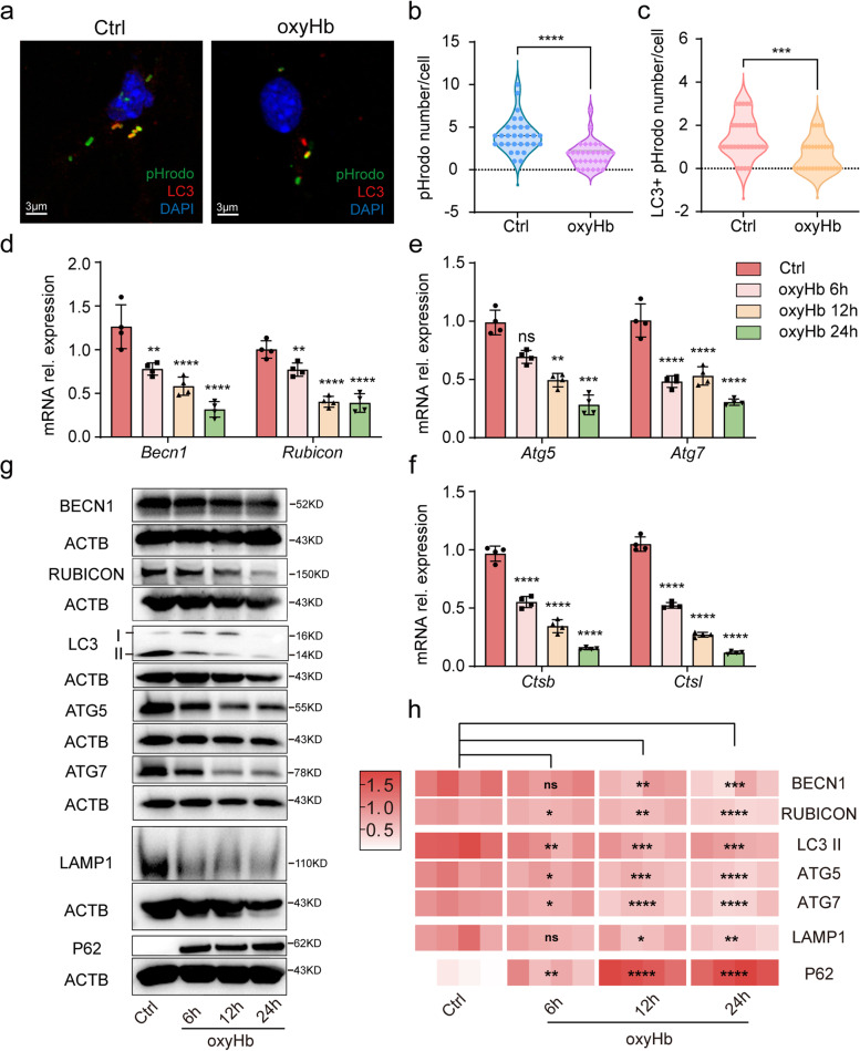
Fig. 2.
Inhibited LAP after oxyHb exposure. Immunofluorescence analysis of pHrodo and LC3 (a) showing the number of particles (b) and LC3 + particles (c) after oxyHb exposure. The relative mRNA expression of Becn1 and Rubicon (d). The relative mRNA expression of Atg5 and Atg7 (e). The relative mRNA expression of Ctsb and Ctsl (f). Protein bands (g) and semi-quantitative analysis (h) showing the protein expression of BECN1, RUBICON, LC3 II, ATG5, ATG7, LAMP1 and P62. Data are presented as mean ± SEM. ****-P < 0.0001, ***-P < 0.001, **-P < 0.01, *-P < 0.05; ns, non-significant. Scale bar: 3 μm

