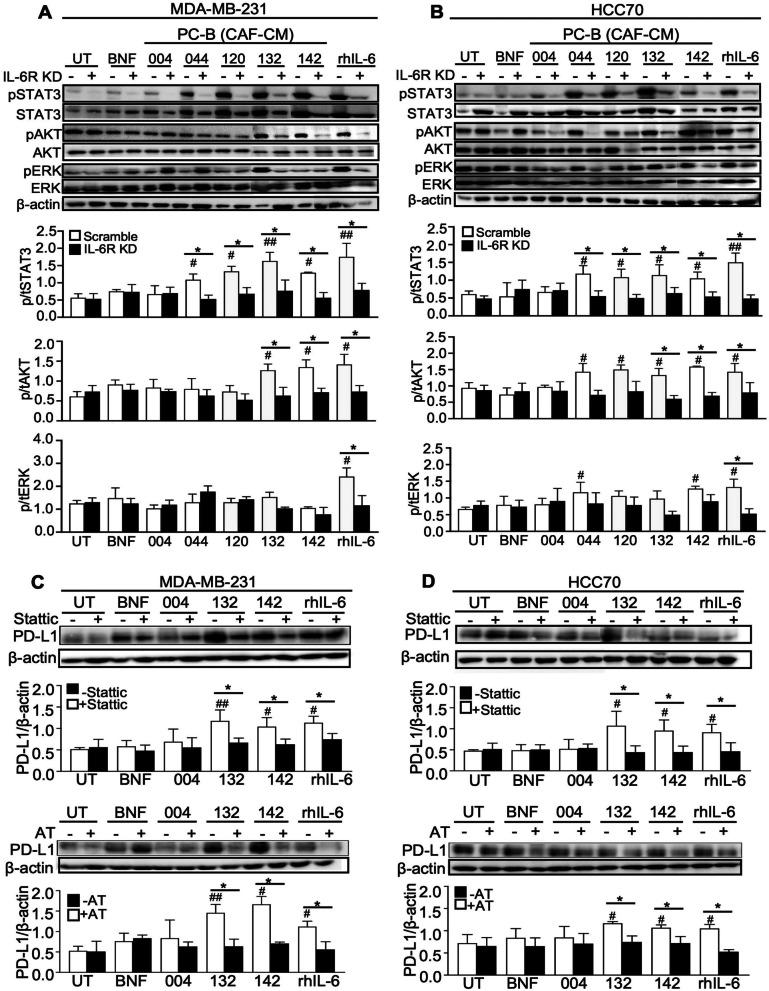
Fig. 4.
Effect of CAF-derived IL-6 on PD-L1 expression through STAT3 and AKT pathways and the blockage with specific inhibitors. A Scramble and siIL-6R KD cells after pre-treating with CM for 30 h to observe the phospho- and total protein levels of STAT3, AKT, and ERK using Western blot analysis in MDA-MD231 and B HCC70 cell lines. The levels of phosphoproteins were normalized against relative total protein and β-actin. C The level of PD-L1 of MDA-MD231 and D HCC70 following blocking with STAT3 (Stattic) or AKT (AT) inhibitors for 2 h. β-actin was used for semiquantitative analysis. Bar graphs represented as mean ± SD of 3 independent experiments. #P < 0.05, ##P < 0.01 compared to BNF using one-way ANOVA, *P < 0.05 compared within group using Student's t test. UT, untreated cells; BNF, breast normal fibroblast; CAFs, cancer-associated fibroblasts; CM, conditioned-medium; IL-6R KD, IL-6R knocked down; rhIL-6, recombinant human IL-6

