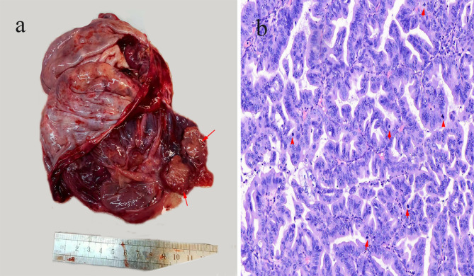
Fig. 2.
(a) Gross examination: The ovarian mass measured 15 × 9 × 4 cm, with a predominantly cystic appearance upon sectioning. The cysts were filled with turbid mucus, and their walls were largely smooth, with occasional surface irregularities and a pink nodular excrescence visible internally (red arrow). (b) Microscopic examination: The glands, exhibiting papillary and cribriform shapes, showed expansile growth with anastomosing architecture and minimal to absent stroma. The lining of most epithelial cells displayed moderate to severe atypia, with diminished or absent mucinous differentiation and conspicuous mitotic figures (red arrow) (H&E, x400)

