
Figure 5.
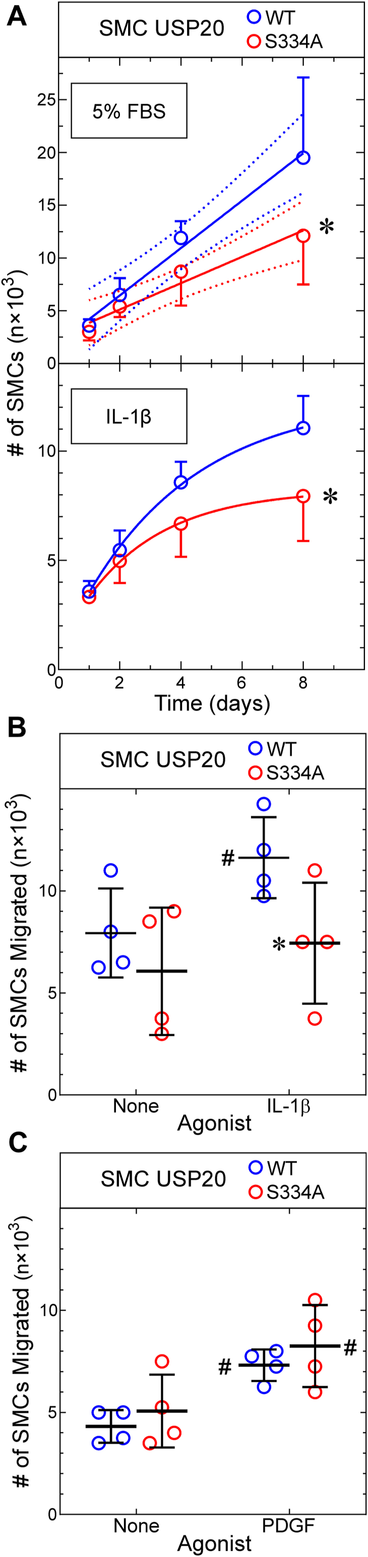
Phosphorylation of USP20 on Ser334 augments SMC proliferation and migration.A, SMCs from WT and USP20-S334A mice were plated at 3 × 10³ SMCs/well in 96-well plates and stimulated to proliferate with either 5% FBS (v/v) (top panel) or IL-1β (20 ng/ml, lower panel), as indicated. Plates were harvested at the indicated times, and SMCs were quantitated as described in Experimental procedures. Shown are means ± SD from three independently isolated SMC lines of each genotype. FBS-stimulated growth was analyzed by linear regression (solid lines, R2 = 0.75 and 0.62 for WT and USP20-S334A, respectively). The line slopes were different from 0 (p ≤ 10−4); the 95% confidence limits for each line are displayed in dotted lines (top panel). Doubling times calculated from linear regression were 2.8 and 4.1 days for WT and USP20-S334A SMCs, respectively (starting at 2 days). IL-β-stimulated growth was fitted by nonlinear regression to an exponential plateau model, and curves were compared by the extra sum-of-squares F test. Compared with WT SMCs: ∗p < 0.03. B and C, SMCs from WT and USP20-S334A mice were plated on Transwell membranes (5 × 10⁴ SMCs/membrane); migration was evoked with 20 ng/ml of IL-1β (B) or 25 ng/ml PDGF-BB (C) as described in Experimental procedures. The number of SMCs migrated to the bottom surface of the membrane is plotted for four experiments with independently isolated SMCs of each genotype, along with means ± SD. Compared with cognate unstimulated SMCs: #p < 0.006; compared with WT: ∗p < 0.025 (paired t tests). IL-1, interleukin-1; SMC, smooth muscle cell; USP20, ubiquitin-specific peptidase 20.