
Figure 7. AlphaFold models of the protein complexes with D/E-PARylation sites in their interfaces.
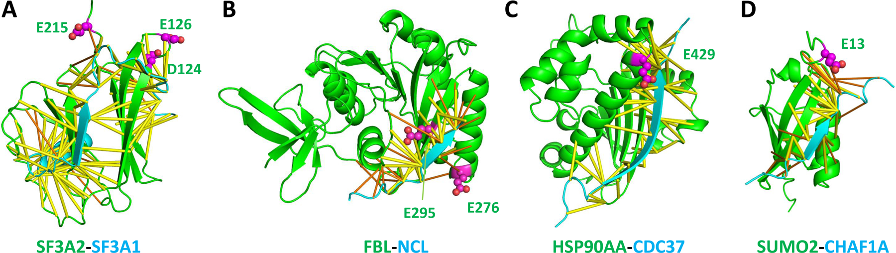
(A) SF3A2 and SF3A1 of the SF3a spliceosome complex. (B) FBL and NCL. (C) HSP90AA and CDC37. (D). SUMO2 and CHAF1A. Predicted contacts and D/E-PARylation sites are shown in the same way as in Figure 3.