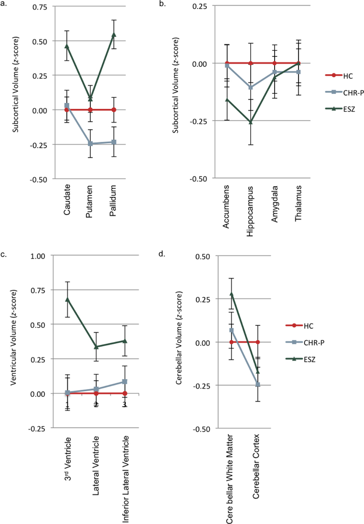
Figure 3.
Subcortical/ventricular/cerebellar volume profiles of healthy control (HC), clinical high-risk (CHR), and early illness schizophrenia (ESZ) groups. a) All groups were nonparallel and had significant level differences. In 2-group follow-up comparisons, HC vs. ESZ and CHR-P vs. ESZ were nonparallel and had significant level differences. b) All groups were parallel and had no significant level differences. c) All groups were parallel and had significant level differences for ventricular volume. ESZ group had larger ventricular volumes relative to HC and CHR-P groups. d) All groups were nonparallel and had no significant level differences. In 2-group follow-up comparisons, HC vs. ESZ and HC vs. CHR were nonparallel.

