
Figure 1.
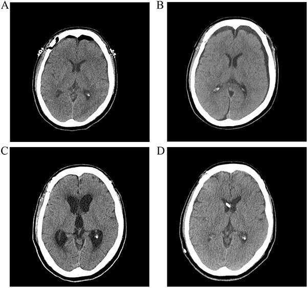
(A) Postoperative CT scan showing no subarachnoid or intraventricular hemorrhages. (B) Bilateral subdural fluid collection on postoperative day 13 CT scan. (C) Increased ventricle size by day 26. The patients developed symptoms of hydrocephalus. (D) A ventriculoperitoneal shunt was placed on day 47. Ventricle size decreased and clinical signs of hydrocephalus improved.