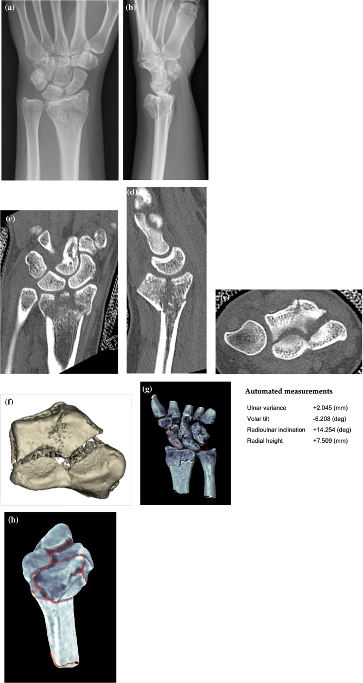
Fig. 2.
a, b Dorsopalmar and lateral projection of radiographs of a 43-year-old female who sustained an intraarticular distal radius fracture by falling on her extended left (adominant) wrist. c–e Coronar, sagittal and axial view of computed tomography images of the wrist of the same patient showing the fracture dislocation and multiple intraarticular fragments. f Three-dimensional reconstruction of the computed tomography images showing the intra-articular fragments and gaps in the articular surface. g Automated measurements conducted by the Bonelogic® 2 software (Disior Ltd, Helsinki, Finland). h Screenshot of the same software conducting automated fracture line detection

