
Fig. 3.
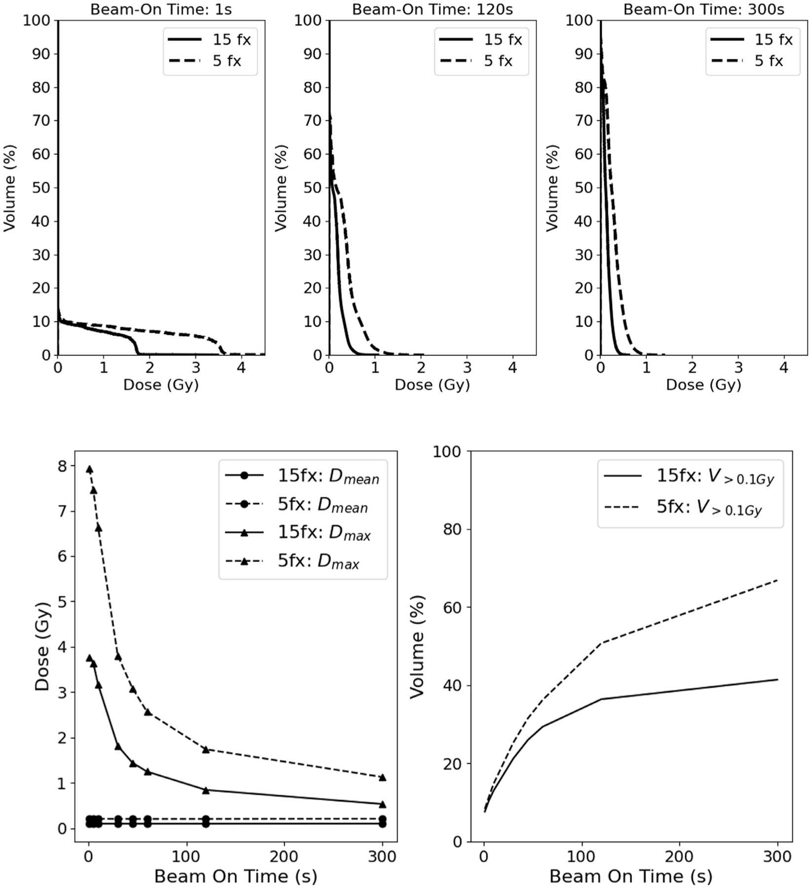
Comparison of the cumulative per fraction bDVH for a 1-second (A, top left), 120-second (B, top middle), and 300-second (C, top right) beam-on time for a sample patient and quantitative comparison of bDVH mean dose, Dmean (D, bottom left), maximum dose, Dmax (D, bottom left), and volume of lymphocytes receiving greater than 0.1 Gy, V>0.1Gy (E, bottom right) taking the mean across all patients between conventional and hypofractionated regimens. Abbreviation: bDVH = bloodstream dose-volume histograms.