
Figure 5: Recurrent PPP1CB-ALK and novel LRRFIP1-ALK fusions are oncogenic drivers in vitro and in vivo.
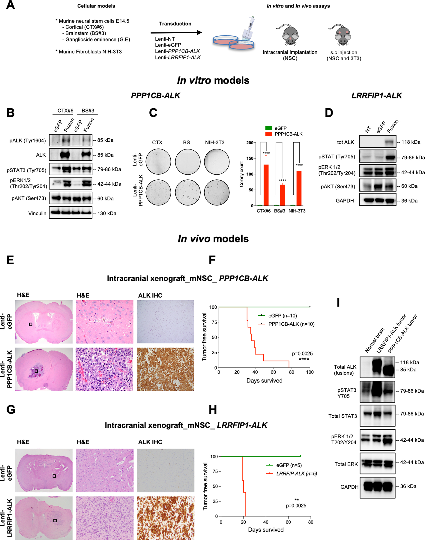
A. Overview of PPP1CB-ALK assessments in vitro and in vivo using murine fibroblasts and mouse NSCs isolated from cortex (CTX), brainstem (BS) and ganglioside eminence (G.E).
B. Representative western blot of PPP1CB-ALK expression and ALK Signaling Pathways in murine neural stem cells (mNSC) isolated from cortex (CTX) and brainstem (BS). Vinculin was used as a loading control.
C. Assessment of the anchorage-independent growth ability of murine NSC and murine fibroblasts expressing PPP1CB-ALK fusion. Quantification of colony formation using CellProfiler. Values represent colony counts ± s.d. The mean of three independent replicates is shown. Error bars show standard error of the mean. Significance between eGFP and PPP1CB-ALK cells is determined by the Mann-Whitney test. ****P < 0.01.
D. Representative western blot of LRRFIP1-ALK expression and ALK Signaling Pathways in murine neural stem cells (mNSC) isolated from cortex (CTX). GAPDH was used as a loading control.
E. H&E and ALK IHC staining of representative mouse brains injected with NSC eGFP (upper row) compared to mNSC PPP1CB-ALK brains (bottom row). H&E boxed areas shown at higher magnification in center.
F. Kaplan-Meier survival curves of SCID mice injected intracranially with mNSC CTX-PPP1CB-ALK (red, n = 10) or mNSC CTX-eGFP (green, n = 10).
G. H&E and ALK IHC staining of representative mouse brains injected with mNSC eGFP (upper row) compared to mNSC LRRFIP1-ALK grafted brains (bottom row). H&E boxed areas shown at higher magnification in center.
H. Kaplan-Meier survival curves of SCID mice injected intracranially with mNSC CTX-PPP1CB-ALK (red, n = 5) or mNSC CTX-eGFP (green, n = 5).
I. Western blot of PPP1CB-ALK and LRRFIP1-ALK tumor lysates from mouse intracranial tumors compared to unrelated normal brain control showing ALK activation effects on STAT and ERK signaling. GAPDH was used as a loading control.