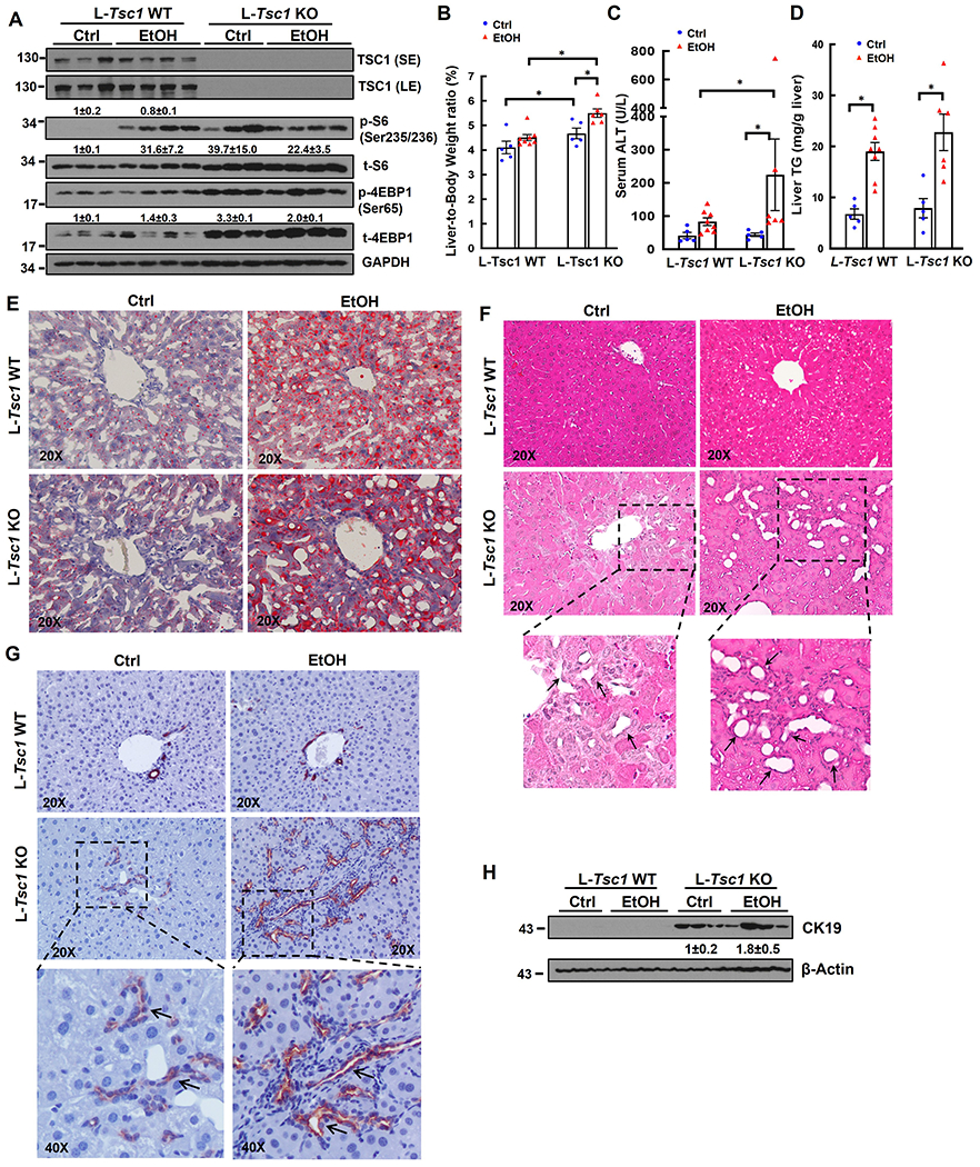
Figure 2. Loss of hepatic TSC1 increases mTORC1 activity and promotes hepatomegaly, ductular reaction and liver injury in EtOH-fed mouse livers.

Male Tsc1 WT and L-Tsc1 KO mice were fed with Gao-binge alcohol. (A) Total liver lysates were subjected to western blot analysis. Data are densitometry analysis and presented as means ± SE (n= 3-4). (B) Liver weight versus body weight ratio, (C) Serum alanine aminotransferase (ALT) levels & (D) hepatic triglyceride (TG) levels were analysis. Data are means ± SE (n= 5-10). *p<0.05; One-Way ANOVA analysis with Bonferroni post hoc test. Representative images of Oil Red O (E), H&E staining (F) and IHC staining of CK19 (G). Arrows denote DR or CK19 positive DR. (H) Total liver lysates were subjected to western blot analysis for CK19.
