
Figure 2: Immature and mature SSC niche.
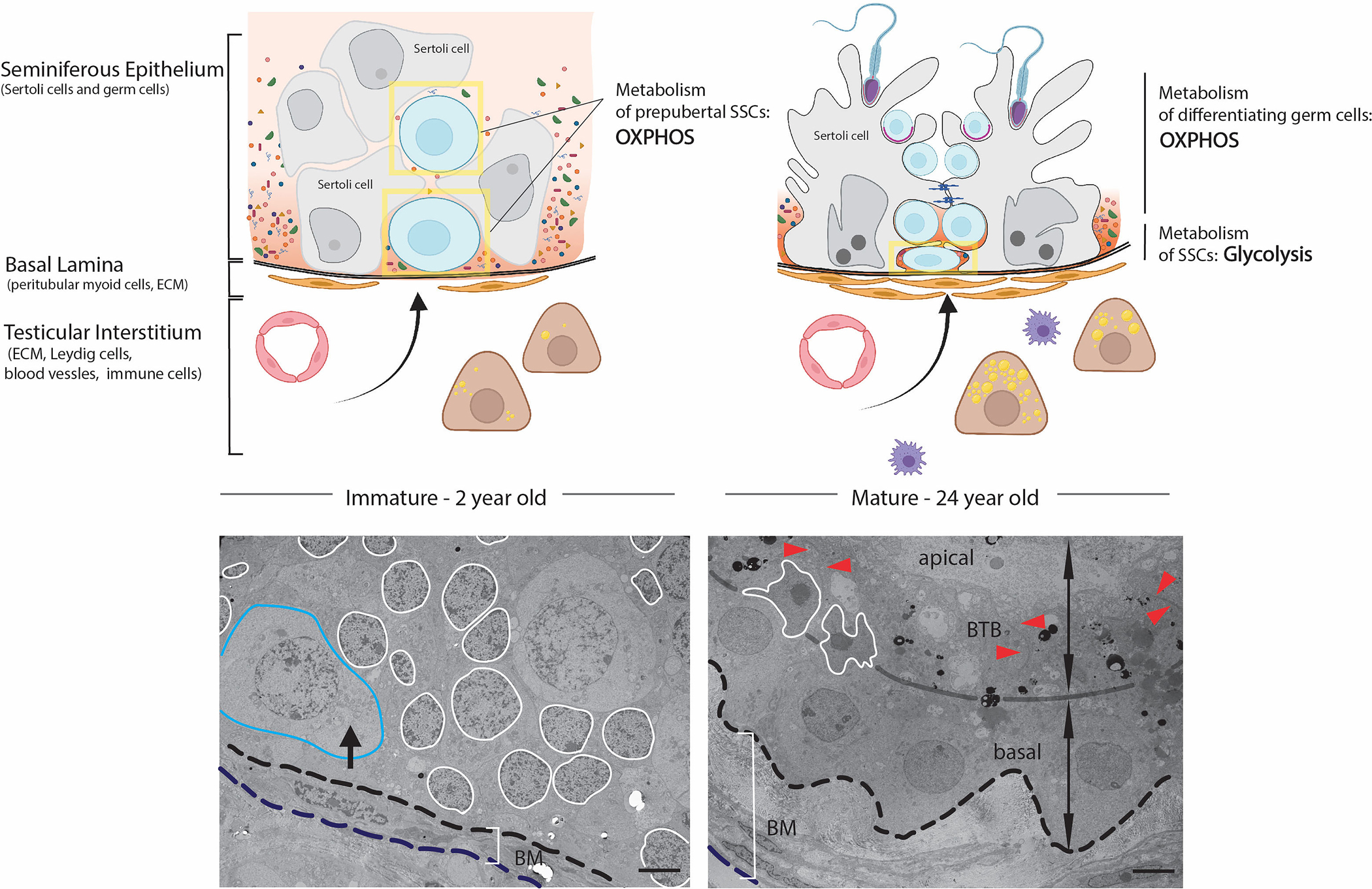
Left: Proliferating Sertoli cells form the primitive niche structure for prepubertal SSCs (in yellow boxes), with a simple basal lamina surrounded undifferentiated peritubular myoid cells and Leydig cells. Blood-derived molecules can freely diffuse through the cords. Immature SSCs are large circular cells and can be found at the basement membrane and occasionally central. Right: In the adult seminiferous epithelium, mature Sertoli cells and blood-testis barrier block diffusion of blood-derived molecules and create a specific composition of metabolites and growth factors to subsidize the anaerobic metabolism supporting adult SSC circuits. A thickened basal lamina and several layers of differentiated peritubular myoid cells together with differentiated testosterone producing Leydig cells and increased numbers of macrophages contribute to the specific microenvironment of the SSC niche. Adult SSCs are flat, half-moon shape and reside close to the basement membrane. TEM of 2-year (left) and 24-year-old (right) human testis: blue outlines prepubertal SSC, white outlines the Sertoli cell nucleus, black arrow shows perinuclear accumulated mitochondria, BM identifies basement membrane, red arrowhead identifies the BTB. Scale bar = 5μm (Figure modified from38).