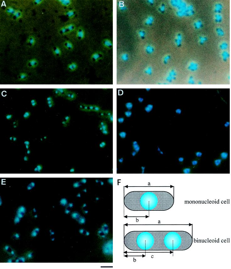
FIG. 1.
Images of M. capricolum cells observed by phase-combined fluorescence microscopy. Cells were fixed, stained with DAPI, and photographed. (A) Culture was grown to an OD600 of 0.05 in the standard medium. (B) Culture was grown to an OD600 of 0.4 without nucleoside supplementation. (C and D) Cells grown to an OD600 of 0.05 in the standard medium were transferred to a medium without supplementation for lipid synthesis and incubated for 1 and 2 h, respectively. (E) Cells grown to an OD600 of 0.05 in the standard medium were incubated with chloramphenicol at 37°C for 1.5 h. Bar, 2 μm. (F) Schematic illustration of M. capricolum cells. a, cell length; b and c, nucleoid positions. Internucleoid distance was calculated by subtraction of b from c.

