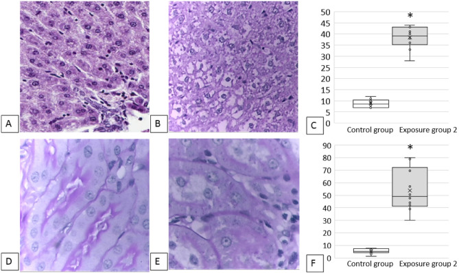
Figure 4.
Results of a histological examination of the experimental animals: (A) liver, control group: normal structure of liver, 100 × magnification; (B) liver, exposure group 2: pronounced dystrophic and necrobiotic changes in hepatocytes, 100 × magnification; (C) the increased number of prokaryotic hepatocytes, %, * p < 0.05 compared with controls; (D) kidneys, control group: the epithelium of convoluted tubules of kidneys with a clear PAS-positive brush border on the apical edge, the cytoplasm is homogeneous, the nuclei are well visualized, the lumens of the tubules are not dilated, 100 × magnification; (E): kidneys, exposure group 2: foci of destruction of the PAS-positive brush border of the tubular epithelium, dystrophic changes in the cytoplasm, tubular luminal dilation, 100 × magnification; (F) loss of the brush border of the renal tubular epithelium, %, * p < 0.05 compared with controls.

