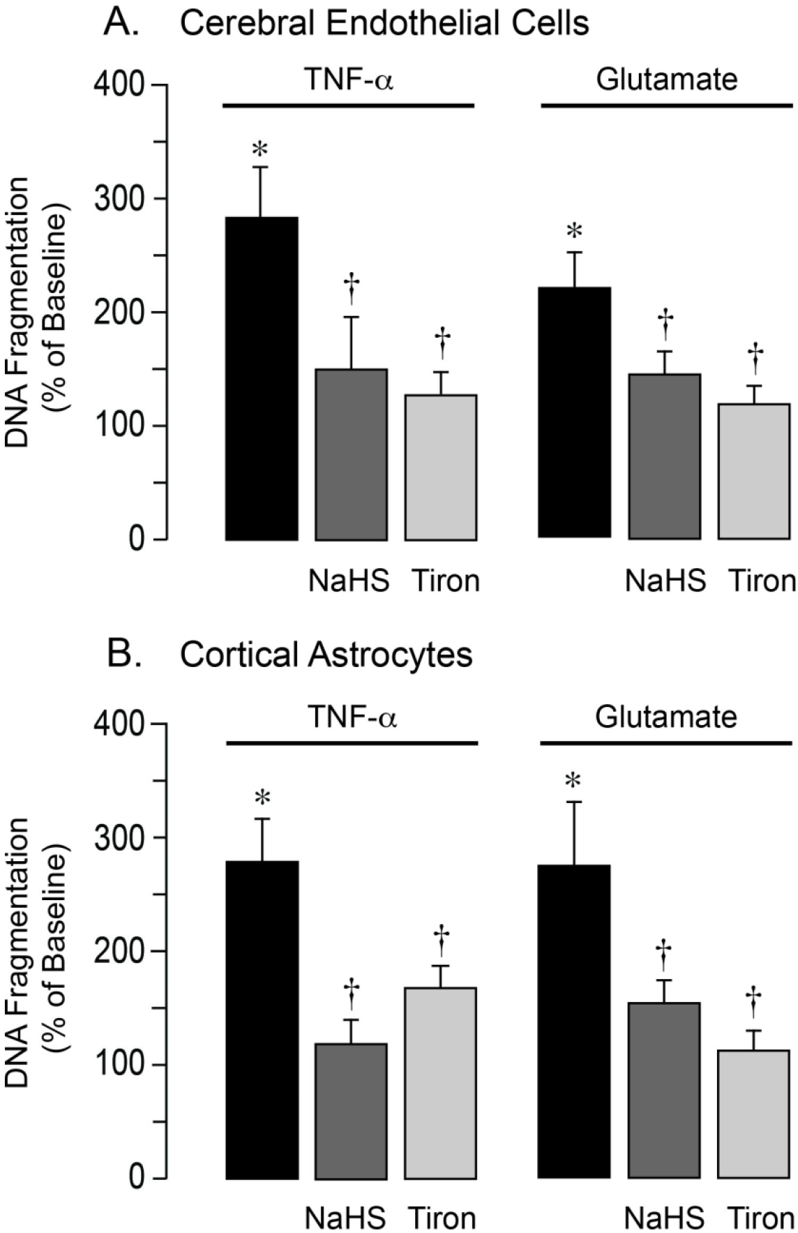
Figure 6.

NaHS prevents apoptosis caused by oxidative stress in cerebral microvascular endothelial cells and cortical astrocytes. Cultured primary cerebral microvascular endothelial cells (A) and cortical astrocytes (B) from newborn pigs were treated with TNF-α (30 ng/ml) or glutamate (2 mM) for 3–5 h in the absence or presence of NaHS (20 μM) or the superoxide scavenger Tiron (1 mM). DNA fragmentation, a key event of apoptosis, was detected by ELISA. Data represent the average of 5 independent experiments. Values are means ± SD. *P < 0.05 compared with the baseline value. †P < 0.05 compared with TNF-α or glutamate alone.
