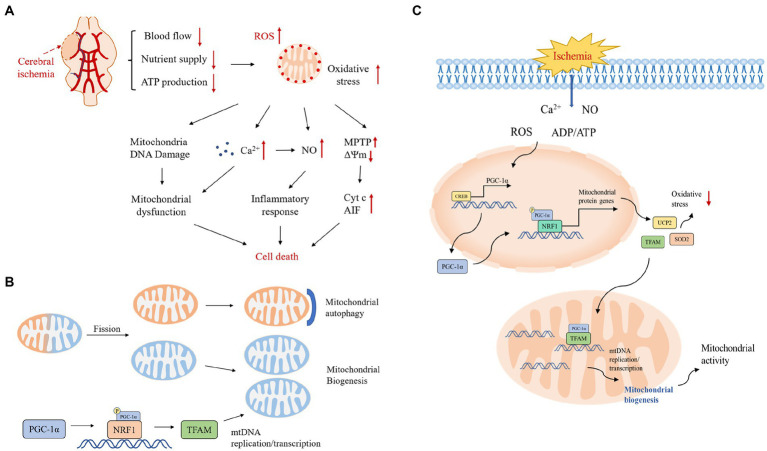
Figure 1.
(A) Pathological mechanisms in the cerebral ischemic cascade response. Excessive production of reactive oxygen species after cerebral ischemia activates various downstream pathological processes, excessive Ca2+ influx, mitochondrial DNA damage leading to mitochondrial dysfunction, activation of inflammatory factors inducing an inflammatory response, and under stressful conditions, transient opening of MPTP in the mitochondrial inner membrane leading to collapse of the mitochondrial transmembrane potential and triggering the release of Cyt c and other pro-apoptotic molecules, which together initiate the apoptotic cascade reaction. ΔΨm, mitochondrial transmembrane potential; Cyt c, cytochrome c; NO, nitric oxide. (B) Mitochondrial quality control mechanisms. Mitochondrial quality control includes the dynamic balance of mitochondrial autophagy, biogenesis, fusion and fission. Mitochondrial autophagy and biogenesis are regulated in a coordinated manner to replace damaged mitochondria during periods of high mitochondrial turnover. When mitochondrial function is dysfunctional under hypoxia, unhealthy components of mitochondria will fission from the healthy mitochondrial network and then degrade to fragments through autophagy; the remaining healthy mitochondrial network can grow and divide through biogenesis. (C) Protective mechanisms of PGC-1α during ischemia-induced stress and a central role in mitochondrial biogenesis. The stress-inducible molecules ROS, Ca2+, ADP/ATP and NO promote the expression of PGC-1α, which upregulates the expression of antioxidant proteins and increases mitochondrial biogenesis to protect neurons from oxidative stress and promote neuronal survival.

