
FIGURE 3.
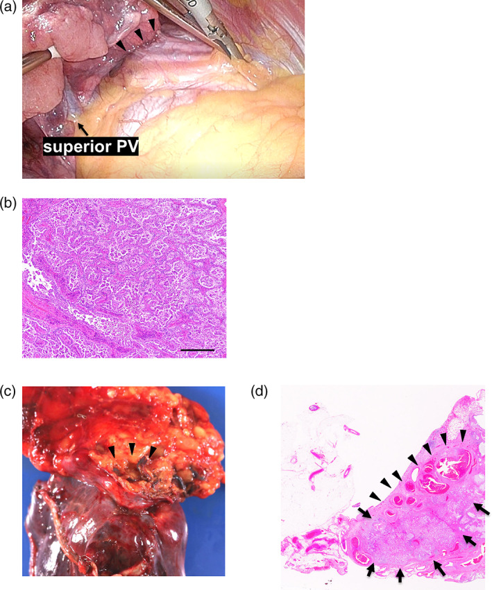
(a) Thoracoscopic findings showed that the right upper lobe was adherent to the anterior mediastinum. PV, pulmonary vein. (b) Microscopic findings of papillary adenocarcinoma. Scale bar, 200 μm. (c) Macroscopic findings in the right upper lobe and anterior mediastinal adipose tissue. A right medial mammary artery fragment can be seen (arrowhead). (d) Pulmonary adenocarcinoma (arrow) and right internal mammary artery inflow (arrowhead) were observed in a loupe view using hematoxylin–eosin staining.