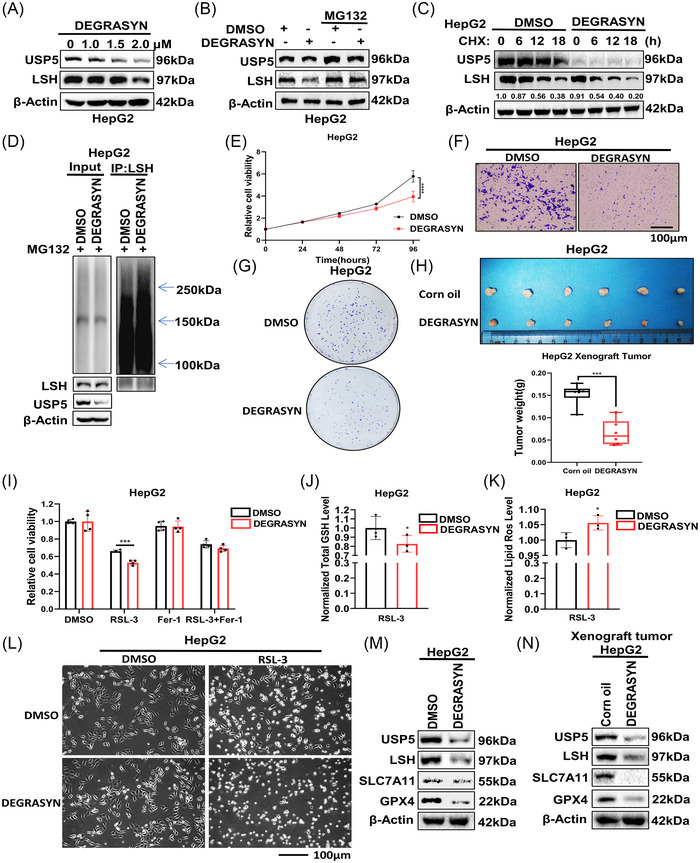
FIGURE 6.

Degrasyn promotes ferroptosis to inhibit tumor progression via targeting of ubiquitin‐specific protease 5 (USP5). (A) Western blot was used to detect the lymphoid‐specific helicase (LSH) expression in HepG2 cells treated with degrasyn at different concentrations. (B) HepG2 cells treated with degrasyn were treated with or without MG132 (20 μM, 12 h), then western blot was used to test the protein level of LSH. (C) HepG2 cells treated with degrasyn were treated with cycloheximide (CHX, 10 mg/mL) for the indicated time followed by WB. (D) Before being collected, degrasyn‐treated HepG2 cells were given MG132 (20 μM, 12 h) treatment. Anti‐LSH was used to immunoprecipitate LSH, and anti‐Ub was used for immunoblotting. (E–G) The cell counting kit‐8 (CCK8) assay (E), transwell assay (F), and colony formation assay (G) of HepG2 cells treated with DMSO or degrasyn. Scale bars, 100 μm. (H) The parental HepG2 cells were transplanted on nude mice treated with corn oil or degrasyn intraperitoneally (25 mg/kg, 3 times/week) (n = 6 mice per group). (I) CCK8 assays were used to analyze the responses of HepG2 cells treated with degrasyn to Fer‐1 (10 μM, 24 h) and RSL‐3 (10 μM, 24 h). (J and K) The levels of total GSH (J) and lipid ROS (K) were analyzed in HepG2 cells treated with degrasyn. (L) Representative phase‐contrast images of degrasyn‐treated HepG2 cells treated with RSL‐3 (10 μM, 24 h). (M and N) Western blot was used to detect LSH and ferroptosis‐related proteins in LM3 cells degrasyn‐treated HepG2 cells (M) and xenograft tumor samples (N). *p < 0.05, ***p < 0.001, and ****p < 0.0001.
