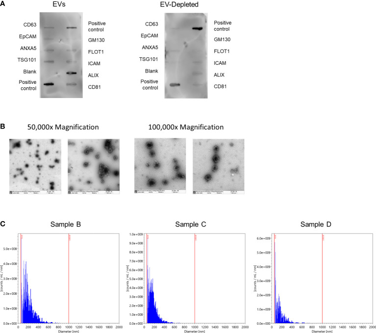
Figure 2.
Characterization of human milk EVs in representative samples. (A) Representative results from Exo-Check Array demonstrating exosome-related protein expression in a pooled sample of three human milk EV samples (left), and the corresponding EV-depleted samples (right), where the darkness of each line indicates the presence of the indicated protein. GM130: Cis-golgi matrix protein, FLOT1: Flotillin-1, ICAM1: Intracellular adhesion molecule 1, ALIX: Programmed cell death 6 interacting protein (PDCD6IP), CD81: Tetraspanin, CD63: Tetraspanin, EpCam: Epithelial cell adhesion molecule, ANXA5: Annexin A5, TSG101: Tumor susceptibility gene 101. (B) Representative transmission electron microscopy images at 50,000x (left) and 100,000x magnification. EVs are the larger dots (electron rich areas), surrounded by lighter membranes. (C) Histograms for the number of particles per milliliter of EVs by particle diameter with bins logarithmically scaled.

