
Figure 2. Transcriptional activation of DR5 by p73 upon CDKI-induced Thr86 dephosphorylation and nuclear translocation.
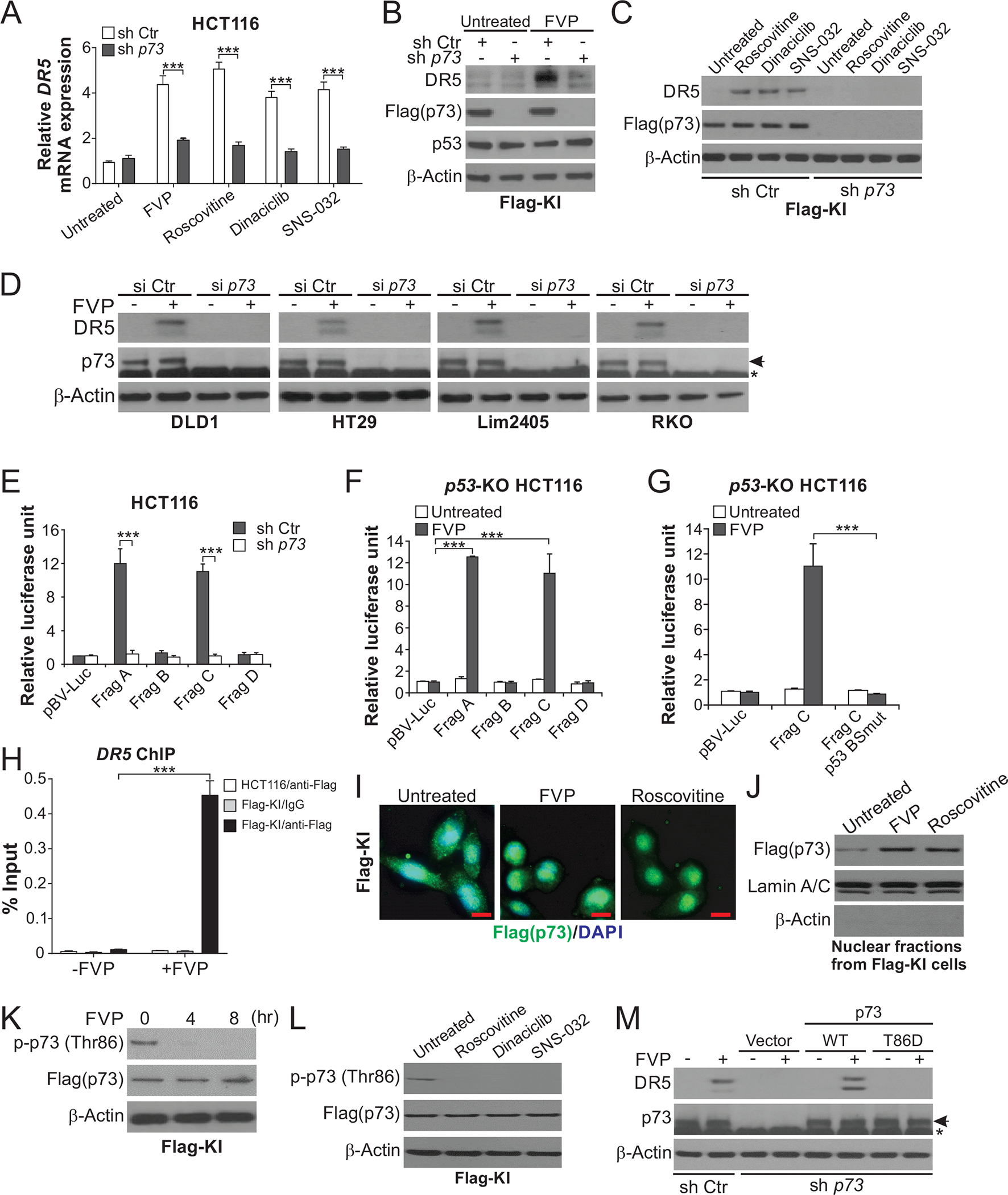
(A) HCT116 cells with stable p73 KD by shRNA (sh p73) or control scrambled shRNA (sh Ctr) were treated with 50 nM FVP, 1 μM roscovitine, 100 nM dinaciclib, or 100 nM SNS-032 for 24 hours. DR5 mRNA expression was analyzed by real-time RT-PCR. (B), (C) Western blotting of indicated proteins in sh p73 and sh Ctr Flag-KI cells treated with CDKIs as in (A). (D) Western blotting of DR5 in indicated CRC cell lines transfected with p73 siRNA (si p73) or control scrambled siRNA (si Ctr) and treated with 50 nM FVP for 24 hours. Arrows indicate p73 and stars indicate nonspecific bands. (E)-(G) Sh p73, sh Ctr, or p53-KO HCT116 cells were transfected with DR5 promoter luciferase reporters and then treated with 50 nM FVP for 24 hours. Reporter activities were measured and normalized to the untreated control samples. In (E) and (F), reporters contain DNA fragments (Frag A-D) covering different regions of the DR5 promoter, among which Frag A and Frag C contain the p53 binding site (Fig. S2F). In (G), reporters contain Frag C with WT or mutant p53 binding site (p53 BSmut) (Fig. S2F). (H) Parental and Flag-KI HCT116 cells with or without treatment with 50 nM FVP for 24 hours were analyzed for the binding of Flag-tagged p73 to the DR5 promoter by chromatin immunoprecipitation (ChIP) using anti-Flag-conjugated beads with IgG as negative control, followed by quantitative real-time PCR analysis of the DR5 promoter region covering the p53 binding site. (I), (J) Flag-KI HCT116 cells were treated with 50 nM FVP or 1 μM roscovitine. Flag-tagged p73 nuclear translocation was analyzed by (I) flag immunofluorescence (green) at 4 hours with DAPI (blue) for nuclear counterstaining (scale bars: 10 μm), and (J) Western blotting of nuclear fractions isolated from cells treated for 24 hours with Lamin A/C (nuclear) and β-actin (cytoplasmic) as loading and fractionation controls. (K), (L) Western blotting of indicated proteins in Flag-KI HCT116 cells treated with (K) 50 nM FVP at indicated time points, or (L) 1 μM roscovitine, 100 nM dinaciclib, or 100 nM SNS-032 for 8 hours. (M) Western blotting of indicated proteins in sh Ctr and sh p73 HCT116 cells transfected with indicated p73 expression constructs and treated with 50 nM FVP for 24 hours. In (A) and (E)-(H), values were expressed as means ± SD of three independent experiments. ***, P < 0.001.