
Figure 4. DR5 is required for apoptosis induced by CDKI/5-FU combinations in CRC cells.
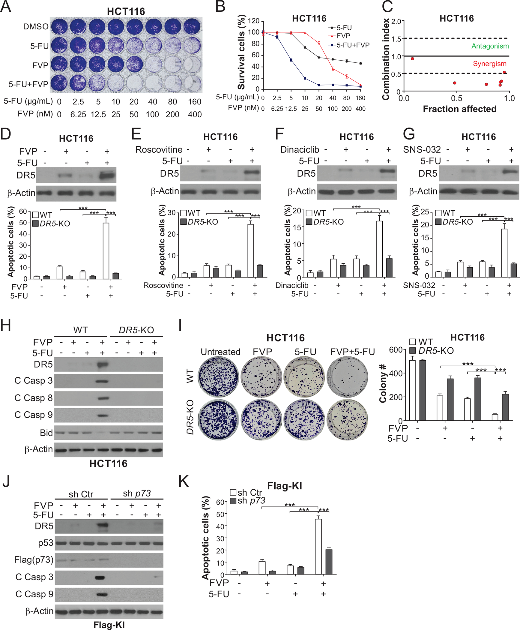
(A)-(C) HCT116 cells treated with FVP alone or in combination with 5-FU at indicated concentrations for 48 hours were analyzed by (A) crystal violet staining of attached cells, (B) MTS analysis of viability, and (C) combination indexes calculated by CompuSyn. (D)-(G) HCT116 cells were treated with 15 μg/mL 5-FU +/− (D) 20 nM FVP, (E) 1 μM roscovitine, (F) 100 nM dinaciclib, or (G) 100 nM SNS-032 for 24 hours. Upper panels: Western blotting of indicated proteins; lower panels: analysis of apoptosis by counting cells with condensed and fragmented nuclei after nuclear staining with Hoechst 33258. (H), (I) WT and DR5-KO HCT116 cells were treated with 20 nM FVP, 15 μg/mL 5-FU, or their combination for 24 hours. (H) Western blotting of indicated proteins. (I) Analysis of long-term cell viability by colony formation assay, which was done by seeding an equal number of treated cells in 6-well plates and staining attached cells with crystal violet on day 14. Left, representative pictures of colonies; right, quantification of colony numbers. (J), (K) Flag-KI HCT116 cells with sh p73 or sh Ctr were treated with 20 nM FVP, 15 μg/mL 5-FU, or their combination for 24 hours. (J) Western blotting of indicated proteins. (K) Analysis of apoptosis by nuclear fragmentation as in (D)-(G). In (B), (D)-(G), (I), and (K), values were expressed as means ± SD of three independent experiments. ***, P < 0.001.